A Few Sleep Tips From Us To You

276

Your mattress and the position you sleep in may affect your spine.

✓ Choose a mattress that provides medium or firm support, such as a traditional coil spring or adjustable airbed. Avoid waterbeds, thick pillow tops and soft, sagging mattresses.
✓ Always sleep on your back with a pillow either underneath your knees or on your side with a pillow between your knees. Avoid sleeping on your stomach.
✓ Keep your neck and back covered while sleeping to avoid drafts that could cause potential muscle spasms.

Here are a couple of tips to help you get in and out of bed more comfortably:
✓ To lie down: Sit on the edge of the bed, pull your arms to your sides and tilt your body into the bed, maintaining the bend of your knees at 45 degrees. Finally, bring your feet into in a lying position or roll onto your back.
✓ To get up: From a side-lying position with your knees bent, push your body upright into a sitting position, swinging your legs over the edge of the bed as you rise.

If you find that you wake up sore then you may be suffering from any number of conditions that get worse overnight.

Workstation Ergonomics

262

Ergonomics is the science of adjusting your workstation to minimize strain in the following ways:

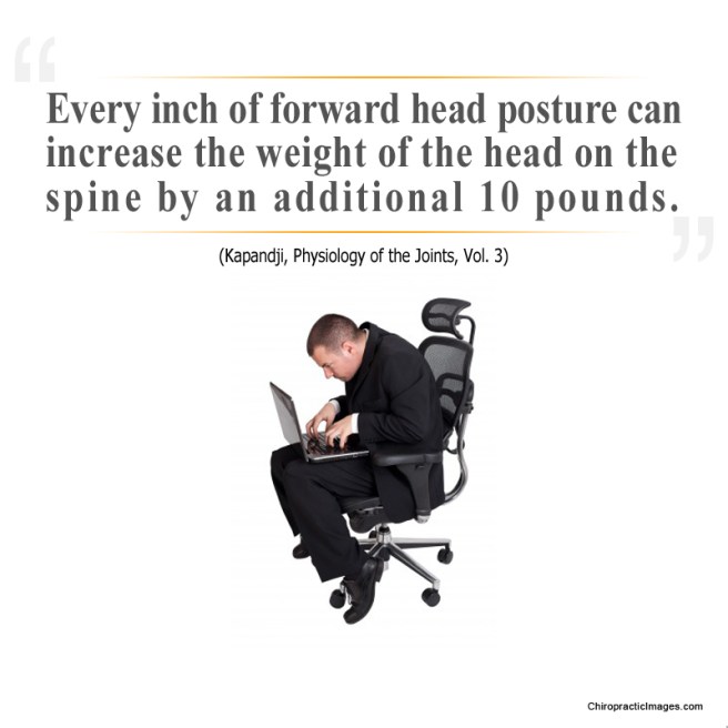
✓ Maintain proper body position and alignment while sitting at your desk – Hips, knees and elbows at 90 degrees, shoulders relaxed, feet flat on floor or footrest.
✓ Wrists should not be bent while at the keyboard. Forearms and wrists should not be leaning on a hard edge.
✓ Use audio equipment that keeps you from bending your neck (i.e., Bluetooth, speakerphones, headsets).
✓ Monitors should be visible without leaning or straining and the top line of type should be 15 degrees below eye level.
✓ Use a lumber roll for lower back support.
✓ Avoid sitting on anything that would create an imbalance or uneven pressure (like your wallet).
✓ Take a 10-second break every 20 minutes: Micro activities include: standing, walking, or moving your head in a “plus sign” fashion.
✓ Periodically, perform the “Brugger relief position” (See video below) -Position your body at the chair’s edge, feet pointed outward. Weight should be on your legs and your abdomen should be relaxed. Tilt your pelvis forward, lift your sternum, arch your back, drop your arms, and roll out your palms while squeezing your shoulders together. Take a few deep cleansing breaths.

How can I reduce stress on my spine while standing?

STress

To avoid extra stress on your spine while standing:

✓ Avoid high-heeled shoes or boots

✓ Use a footrest measuring 10% of your height

✓ To decrease stress on your back and feet consider leaning on a tall chair.

✓ If excessive standing can’t be avoided, consider shock-absorbent shoes or an anti-fatigue mat.

✓ When transitioning from a sitting workstation to a standing desk, begin gradually by standing 20 minutes per hour and not necessarily in a continuous period. Add an extra 10 minutes per hour each day as long as there is no prolonged stiffness or discomfort.

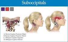
Trigger points in the suboccipitals.

img_0248-1

The suboccipitals are a group or four muscles that attach to the transverse and spinous process of C1 and C2 and the occipital bone.

These muscles provide extension side bending and rotation movements between the occiput and C1 and C2. These muscles are often overloaded due to postural strain. A classic example being sitting in front of a computer all day.

When these muscles are overloaded trigger points can develop. Pain and symptoms of trigger points in the suboccipitals include head pain that penetrates into the skull but is difficult to localize.

Patients are likely to describe the pain as “all over” including the occiput, eye and forehead, but without any clarity. Trigger points in these muscles are often associated with tension head aches

.

Our Exercise Of The Month: ITB Foam Roller

ITB – Foam Roller
  • Lie on your side with your affected thigh over a foam roll.
  • Lift your shoulders off of the floor with your arm and roll the fleshy part your mid & lower thigh back and forth over the foam roller.
  • Avoid rolling over the bony point of your hip unless specifically directed.
  • Perform this exercise for one minute twice per day or as directed.

Hip or Low Back?

​Lumbar and Pelvis

Technically, the hip is the ball-and-socket joint between the long bone of the thigh and the pelvis; but more often than not, many people will point to a number of different places on their back or pelvis and say, “My hip is killing me” when it’s not really “the hip” at all!
Hip pain can be located in the front (groin area), the side, the back, or in the buttocks. The “classic sign” of hip pain is reproduced most consistently when you try to cross your legs—put your ankle on the far end of the thigh and then push down on your knee. This may feel tight and cause pain in the groin area. For many people, hip pain is also reproduced when they cross their legs and then pull their knee towards the opposite shoulder.
The hip is a VERY strong joint due to the deep receiving cup of the pelvis and the round ball that fits nicely into it. Because it’s a freely moving joint, there is a joint capsule. The capsule is lined with tissue that produces an oily substance that lubricates the joint (called synovial fluid), and when hip pain occurs, this can be caused by a capsulitis (inflammation of the capsule) with a buildup of synovial fluid (called synovitis).
When the smooth, shiny surface of the ball starts to wear thin (which can eventually wear away down to the bone), that’s a condition called “osteoarthritis.” This occurs over time for many and may eventually result in the need for a hip replacement. This usually isn’t needed until an individual is in their late 60s or older (if at all), but for those who injure a hip earlier in life, the “wearing out” process may accelerate and a hip replacement may be needed well before old age.
There are many studies that report low back and hip arthritis often occur together, and differentiating between the two can sometimes be a challenge. For example, pain can radiate from the hip to the knee, which many doctors will diagnose as “sciatic nerve.” But hip pain can present exactly the same, making it hard to determine if it’s low back-generated pain or hip-generated pain.
This is why it’s SO IMPORTANT that your doctor of chiropractic conduct a careful history and examination. There are specific tests that he or she will perform that help determine which of the two is causing the pain. There are times when they may find BOTH problems co-existing together, making it necessary to manage two problems, rather than just one.
There are many mobilization, manipulation, soft tissue therapies, modalities, and exercises available to patients with both hip and low back pain. So if you’re not sure what is bothering you and you don’t know what to do, visit your doctor of chiropractic and he or she will help you through this.

Exercise Of The Month: Pike On A Ball

Pike on a Ball

Begin in a push-up position with your knees resting on an exercise ball. Maintain a neutral spine, and do not allow your back to arch down towards the floor. Roll the ball from your thighs to your ankles by bending your knees and bringing them towards your chest. Slowly return to the start position. Perform three sets of 10 repetitions daily or as directed.

What is Facet Syndrome?

​

As stated in last week’s article, low back pain (LBP) can arise from many different structures. Lumbar facet syndrome is one that involves the facet joint and includes both acute (new) and chronic (old) varieties. The facet joint is synonymous with the zygapophyseal joint, so if you hear that word, don’t let it throw you off! Approximately 45% of patients with chronic low back pain suffer from “facet syndrome” (FS) in which the facets are the low back pain generator.
There are many conditions that give rise to FS. Some of these include the straining of the surrounding joint capsule (the capsule holds the joint securely together), joint hypomobility (reduced motion in the joint), a synovial cyst (similar to a ganglion on the back of the wrist but its located inside the joint), and degeneration (also called osteoarthritis—the wearing out type of arthritis).
Because facet syndrome can accompany other conditions, a doctor of chiropractic must evaluate each patient individually and manage each person appropriately. In “pure” facet syndrome, pain rarely ever passes the level of the knee and does not cause neurological loss (weakness, loss of reflex, etc.). It can create numbness but usually NOT beyond the knee. Pain is usually not worsened by hip movements such as straight leg raise or hip rotation.
The facet joint’s “job” (at least in part) is to limit or guard twisting movements in the upper lumbar/low back region, and the lower lumbar facets are shaped to limit motion when bending forwards and backwards. Facet joints are unique because they are innervated by specific nerves that can be blocked by injecting an anesthetic agent to determine if the facet (and its innervating nerve) is the main source of pain. The surrounding capsule around the facet joint contains mechanoreceptors (cells that detect movement) and nociceptors (cells that detect pain) that fire when the facet joint is compressed/jammed or over-stretched. These nociceptors can become “hypersensitized” (very irritable) when they remain inflamed over time.
In many patients, injury to a facet joint is the result of many microtraumas over a period of time and not one single isolated event. For example, repeatedly bending backwards, twisting, and leaning to one side can stretch the joint capsule and fatigue it until some capsular tissues finally “give” and it inflames which generates pain.
These joints commonly become arthritic with age, which is one reason people over 50-60 years old commonly present with FS. Osteoarthritis results in a narrowing of the joint space and causes a more permanently “jammed” joint. This is one reason many elderly people walk partially bent over—as bending forwards opens the facet joints and “feels good!”
facet joint
The good news is that chiropractic manipulation is a highly effective treatment for facet syndrome, and most patients feel much better within the first or second week of care (often within three to five visits).

What Causes Low Back Pain?

​236

Low back pain (LBP) can arise from disks, nerves, joints, and the surrounding soft tissues.
To simplify the task of determining “What is causing my LBP?” the Quebec Task Force recommends that LBP be divided into three main categories: 1) Mechanical LBP; 2) Nerve root related back pain; and 3) Pathology or fracture. We will address the first two, as they are most commonly managed by chiropractors.
Making the proper diagnosis points your doctor in the right direction regarding treatment. It avoids time wasted by treating an unrelated condition, which runs the risk of increased chances of a poor and/or prolonged recovery. Low back pain is no exception! The “correct” diagnosis allows treatment to be focused and specific so that it will yield the best results.
Mechanical low back pain is the most commonly seen type of back pain, and it encompasses pain that arises from sprains, strains, facet and sacroiliac (SI) syndromes, and more. The main difference between this and nerve root-related LBP is the ABSENCE of a pinched nerve. Hence, pain typically does NOT radiate, and if it does, it rarely goes beyond the knee and normally does not cause weakness in the leg.
The mechanism of injury for both types of LBP can occur when a person does too much, maintains an awkward position for too long, or over bends, lifts, and/or twists. However, LBP can also occur “insidiously” or for seemingly no reason at all. However, in most cases, if one thinks hard enough, they can identify an event or a series of “micro-traumas” extending back in time that may be the “cause” of their current low back pain issues.
Nerve root-related LBP is less common but it is often more severe—as the pain associated with a pinched nerve is often very sharp, can radiate down a leg often to the foot, and cause numbness, tingling, and muscle weakness. The location of the weakness depends on which nerve is pinched. Think of the nerve as a wire to a light and the switch of the nerve is located in the back where it exits the spine. When the switch is turned on (the nerve is pinched), and the “light” turns on—possibly in the outer foot, middle foot, inner foot, or front, back or side of the thigh. In fact, there are seven nerves that innervate or “run” into our leg, so usually, a very specific location “lights up” in the limb.
Determining the cause of your low back pain helps your doctor of chiropractic determine which treatments may work best to alleviate your pain as well as where such treatments can be focused.