Did you know that tennis elbow and golfers elbow pain can be caused by trigger points? Points located in the forearm flexor and extensor muscles can cause pain, stiffness, and weakness in the elbow, forearm, wrist, and hand. Quite often these trigger point symptoms are mistaken for inflammation of the tendons which is what a true tennis/golfers elbow actually is. Failure to address the trigger points can lead to an incomplete recovery .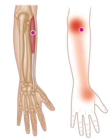
Winnipeg Chiropractor
Trigger point massage
Session Description
A treatment with Bryan is very user friendly. And, no, you don’t have to remove any clothing. However, bringing a t-shirt and a pair of shorts or sweats is recommended.
The first time you come for a treatment you will be asked to fill out a Client History form. Bryan will go over the information you provide, asking for more detail and discussing the type of pain you are having and its location.
The treatment itself involves locating the Trigger Points in the muscle or soft tissue and applying a deep focused pressure to the Point. This will reproduce the pain and the referral pattern that is characteristic of that pain.
The treatment will be uncomfortable at first, but as the Trigger Points release, the pain will decrease. The pressure will always be adjusted to your tolerance level. If, at any time, you feel too uncomfortable you can ask Bryan to ease off a bit.
Depending on your specific problem, Bryan may also use some stretching and / or range-of-motion techniques, as needed.
After treatment, it is usually recommended that the client apply moist heat to the area treate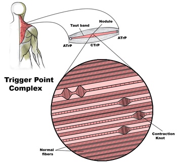
 d.
d.
Problems associated with trigger points
Trigger Points in muscle and other soft tissue are one of the most common causes of a wide variety of pain and dysfunction, including (but not limited to):
• Achy persistent pain
• Severe local pain
• Arm / leg pain
• Back pain
• Radiating pain
• Weakness
• Stiffness
• Pain resulting from a medical condition, such as
– Migraines
– Sciatica
– TMJ dysfunctions
– Arthritis
– Fibromyalgia
– Carpal tunnel syndrome
– Soft tissue injuries
– And more…
Trigger points in the levator scapula
The levator scapula is a muscle located in your neck. It originates on the transverse processes of C1-C4 vertebrae. It inserts on the superior part of the medial border of the scapula. This muscle acts to elevate the scapula and rotate the glenoid fossa downward. At the cervical attachment it acts to rotate the neck to the same side and assists extension. Trigger points will refer pain into the angle of the neck with spillover into the scapula. When this muscle is tight due to trigger points it will restrict neck rotation causing the classic stiff neck. With a forward head position this muscle often becomes stretched and over worked.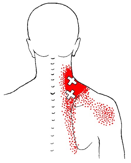
2 Critical Steps to Resolving Ankle Sprains

Efficient treatment of ankle sprains continues well after the pain subsides. While the majority of inversion (lateral) ankle sprains heal relatively quickly, up to 1/3 of patients, continue to note symptoms at one year, and up to 25% report pain, instability, crepitus, weakness, stiffness, or swelling at three years. (1) Re-injury is frequent, with rates reaching almost 75% in sports, like basketball. (2) Successful management of ankle sprains and prevention of re-injury are predicated on a couple of fundamental principles.
Management of ankle inversion sprains requires two steps; each is equally important.
- The first step entails the evaluation and treatment of acute pain.
- The second step involves preventing subsequent sprains – and new research validates the importance of chiropractic care to improve clinical outcomes in these recalcitrant cases.
STEP 1—Move for Pain Relief
Early return to activity for acute inversion sprains is supported by the literature. Exercises and treatments that promote joint motion and early return to weight bearing for acute ankle sprains have proven more effective than immobilization. While grade III sprains (ligament rupture) may require immobilization, grade I and II ankle sprains should forego complete immobilization and instead focus on regaining full range of motion. In fact, early rehab and return to weight bearing will increase ankle range of motion, decrease pain, and reduce swelling sooner than immobilization.
In a study by Linde et al., 150 patients with inversion ankle sprains were treated with early motion and weight bearing. After one month, 90% of the patients treated with early motion and weight bearing demonstrated pain-free gait and 97% had increased work ability. (3) Early mobility exercises would typically include:


These four exercises promote balance and range of motion – specifically dorsiflexion, which is a key contributor to ankle injury. Patients who have lost an average of 11 degrees of dorsiflexion are five times more likely to suffer lateral ankle sprains. (4)
In office care should also include mobilization and manipulation for restoring function. Joint mobilization has been shown to decrease pain, increase dorsiflexion, and improve ankle function. (5) IASTM or transverse friction massage to the affected ligament may help mobilize scar tissue and increase pliability. Myofascial release may help release tightness or adhesions in the gastroc and soleus. (Side note: The FAKTR concept seamlessly incorporates all of these tools to produce top-tier outcomes.)
Knowing when to treat and when to refer is critical. Whitman’s clinical prediction rule identifies four variables to predict the success of manipulation and exercise for the treatment of inversion ankle sprains. (6) The presence of three out of four of the following variables predict greater than a 95% success rate for manual therapy and exercise:
- Symptoms worse when standing
- Symptoms worse in the evening
- Navicular drop greater than 5 mm
- Distal tibiofibular joint hypomobility
STEP 2- Prevent Re-injury
The second step is shorter and easier than the first. The most crucial variable in the successful prevention of future ankle sprains is improving BALANCE. Balance training reduces the incidence of ankle sprains and increases dynamic neuromuscular control, postural sway, and joint position sense in athletes. (7) A study by de Vasconcelos et al. (2018) found that balance training reduced the incidence of ankle sprains by 38% compared with the control group. (7)
Two of the most common exercises used for balance and proprioception include the single-leg stance exercise and Veles. A simple explanation stressing the importance of balance training may be necessary to promote patient compliance.
Finally, encourage your patients start walking “normal” as soon as possible. As evidence-based chiropractors, we need to return patients back to their normal gait as soon as tolerable. Patients with foot and ankle pain will often favor a supinated gait in order to unload the soft tissues of the foot and arch in favor of their bony architecture on the lateral foot. The lateral column of the foot affords stability but at the expense of a very inefficient gait. Over an extended period, these patients may develop a Tailor’s bunion, i.e. 5th metatarsal head bursitis. However, in the case of ankle sprains, a rapid increase in activity may overload the metatarsal fast enough to cause a Jones Fracture. Return to normal gait will minimize these compensations.
Pregnancy Related Low Back Pain

Low back pain during pregnancy is quite common. In fact, between 50-75% of all pregnant women will experience low back pain. The pain is usually caused from rapid changes in weight, posture, gait and hormones.
The average woman gains between 20-40 pounds throughout pregnancy. This weight gain moves your center of gravity forward, causing your pelvis to tilt and your lower back to sway – placing excessive stress on the ligaments, discs, and joints of your spine.
 Pregnancy-related low back pain typically starts between the fifth and seventh month of pregnancy, although a significant portion of women experience pain sooner. Symptoms often begin at the base of your spine and may radiate into your buttock or thigh. Discomfort is often aggravated by prolonged standing, sitting, coughing, or sneezing. Your symptoms may increase throughout the day, and some patients report nighttime pain that disturbs their sleep. The extremes of activity seem to contribute to pregnancy-related low back pain – with increased risk for both “sedentary” and “physically demanding” lifestyles. Patients who have suffered with back pain prior to pregnancy are more than twice as likely to re-develop back pain during pregnancy.
Be sure to tell your doctor if your symptoms include fever, chills, bleeding, spotting, unusual discharge, cramping, sudden onset pelvis pain, light-headedness, shortness of breath, chest pain, headache, calf pain or swelling, decreased fetal movement, or symptoms that radiate beyond your knee.
Unfortunately, pregnancy related low back pain occurs at a time when your medical treatment options are limited. Not surprisingly, over 90% of prenatal health care providers would recommend drug-free treatment, including the type of alternative therapy provided in this office. Studies have shown that chiropractic manipulation provides significant relief of pregnancy-related low back pain. Almost 75% of women undergoing chiropractic care report significant pain reduction with improved ability to function.
Most patients will also benefit from continuing aerobic exercise throughout pregnancy. The US Department of Health and Human Services advises that healthy pregnant women may begin or continue moderate intensity aerobic exercise for at least 150 minutes per week. Women should not begin “vigorous” exercise during pregnancy, but those who were preconditioned to vigorous exercise may continue. Be sure to check with your doctor prior to initiating or increasing any exercise program while you are pregnant.
Be sure to take frequent breaks from prolonged sitting or standing. You may find benefit by using a small foot stool to alternate feet while standing. Sleeping with a pillow between the knees in a side lying posture may help you to rest more comfortably. You should wear shoes with good arch supports. In some cases, your chiropractor may recommend a sacroiliac belt or pelvic support belt to help relieve your pregnancy-related low back pain.
Exercise of the Month – (Resisted Shoulder Retraction)
Resisted Shoulder Retraction
- Secure a piece of elastic resistance tubing to a doorframe.
- Sit or stand with your elbows tucked into your sides bent at 90 degrees, forearms pointing forward.
- Grasp the resistance band and pull it towards you by focusing on pinching your shoulder blades together.
- Return to the start position and repeat three sets of 10 repetitions daily or as directed.
*This exercise may also be performed using a cable row machine or by looping a piece of elastic resistance band over your feet while sitting on the floor with your legs directly in front of you.
Lumbar Radiculopathy? That sounds ridiculous!
Your nervous system is basically a big electrical circuit. Your spinal cord transmits all of the electrical nerve impulses between your brain and lower back. From there, individual nerves emerge from your spine then travel to supply sensation and movement to a specific area of your buttock, legs and/or feet. This allows you to move and feel sensations like touch, heat, cold and pain. Anything that

interferes with this transmission can cause problems.
You have been diagnosed with a “Lumbar Radiculopathy”. This means that one or more of the nerves emerging from your lower back has become irritated or possibly pinched. This often results in pain, numbness or tingling in the specific area of your leg that is supplied by the irritated nerve. The term “Sciatica” is often used to describe this condition, because most (but not all) “lumbar radiculopathies” involve the sciatic nerve which supplies the back & outside of your thigh and calf. Symptoms of a lumbar radiculopathy may vary from a dull ache to a constant severe sharp shooting pain. Your symptoms are likely aggravated by certain positions or movements.
To solve this problem, we will treat the source of your nerve irritation. It is important for you to follow your treatment plan closely and be sure to tell us immediately if you experience any progression of your leg pain, numbness or weakness.
Lumbar Degenerative Spondylolisthesis
Your lumbar spine (low back) is made up of five individual vertebra stacked on top of a bone called the sacrum. To allow for flexibility and movement, there is a cushion or “disc” in between each level. As we age, our discs and joints can wear and become thinner from a process called arthritis. This leads to additional changes, including loosening of the ligaments that hold your vertebra in place.

The term “degenerative lumbar spondylolisthesis” means that one of your vertebra has shifted forward on top of the one below as a result of arthritis and loosening ligaments. The condition usually comes on after age 50 and affects women six times more frequently than men. Degenerative spondylolisthesis occurs most commonly at your second lowest spinal level. (L4-5)
Sometimes, spondylolisthesis develops silently, but most patients report episodes of back pain that have occurred for many years. Patients often report increased pain when standing or when rising from a sitting position. Pain tends to increase throughout the day. If your nerve openings have become narrowed, the nerves may be pinched, and you may experience pain radiating into your legs. Leg symptoms that shift from side to side are characteristic of degenerative spondylolisthesis. Leg pain and tingling are fairly common, but be sure to tell your doctor if you notice more significant symptoms, like leg numbness, heaviness, weakness, loss of bowel or bladder function, or impotence.
Studies have shown no advantage for surgery over conservative care for most cases of degenerative spondylolisthesis. Approximately one-third of patients will experience progression of symptoms over time, and only 10-15% will ever need surgery to correct the problem. Fortunately, the majority of patients will benefit from treatment and exercises to help stabilize their spine.
You will need to perform your exercises consistently for sustained improvement. You should also try to add some type of aerobic exercise to your daily routine. Stationary cycling is a very good choice, and other options include water walking and swimming. Avoid wearing high heels. You may find some benefit for your arthritic symptoms by taking 1500mg of Glucosamine Sulfate each day. Using a hot pack for 10-15 minutes directly over your lower back may provide some benefit.
A Modern Spine Ailment
Some great information from Spine-Health.com and Dr. Steven Shoshany DC
Text neck is the term used to describe the neck pain and damage sustained from looking down at your cell phone, tablet, or other wireless devices too frequently and for too long.

Using a mobile device often can lead to poor posture and symptoms of text neck.
Watch: Text Neck Treatment Video<spanclass=”div-video-link”></spanclass=”div-video-link”>
And it seems increasingly common. Recently, a patient came in to my practice complaining of severe upper back pain. He woke up and was experiencing severe, acute, upper back muscle strain. I told him I believe the pain is due to the hours he was spending hunched over his cell phone. Diagnosis: Text neck.
Of course, this posture of bending your neck to look down does not occur only when texting. For years, we’ve all looked down to read. The problem with texting is that it adds one more activity that causes us to look down—and people tend to do it for much longer periods. It is especially concerning because young, growing children could possibly cause permanent damage to their cervical spines that could lead to lifelong neck pain.
See Cervical Spine Anatomy and Neck Pain
What are the symptoms associated with text neck?
Text neck most commonly causes neck pain and soreness. In addition, looking down at your cell phone too much each day can lead to:
-
- Upper back pain ranging from a chronic, nagging pain to sharp, severe upper back muscle spasms.
- Shoulder pain and tightness, possibly resulting in painful shoulder muscle spasm.
- If a cervical nerve becomes pinched, pain and possibly neurological symptoms can radiate down your arm and into your hand.
See What Is Cervical Radiculopathy?
I believe, as some studies suggest, text neck may possibly lead to chronic problems due to early onset of arthritis in the neck.
See Facet Joint Osteoarthritis
How common is text neck?
A recent study shows that 79% of the population between the ages 18 and 44 have their cell phones with them almost all the time—with only 2 hours of their waking day spent without their cell phone on hand.1
How is text neck treated?
First, prevention is key. Here are several pieces of advice for preventing the development or advancement of text neck:
-
- Hold your cell phone at eye level as much as possible. The same holds true for all screens—laptops and tablets should also be positioned so the screen is at eye level and you don’t have to bend your head forward or look down to view it.
See Ten Tips for Improving Posture and Ergonomics
-
- Take frequent breaks from your phone and laptop throughout the day. For example, set a timer or alarm that reminds you to get up and walk around every 20 to 30 minutes.
- If you work in an office, make sure your screen is set up so that when you look at it you are looking forward, with your head positioned squarely in line with your shoulders and spine.
See Ergonomics of the Office and Workplace: An Overview
The bottom line is to avoid looking down with your head bent forward for extended periods throughout the day. Spend a whole day being mindful of your posture—is your head bent forward when you drive? When you watch TV? Any prolonged period when your head is looking down is a time when you are putting excessive strain on your neck.
See Office Chair, Posture, and Driving Ergonomics

Keeping the neck straight and your phone at eye level can help prevent text neck.
Watch: Neck Strains and Sprains Video
Next, rehabilitation is important.

-
- Many people don’t know this, but you need to have strong core muscles—the abdominal and lower back muscles—to support your upper body, including your neck. Your core muscles usually do not get enough exercise during normal daily activities, so you need to do specific exercises to target these muscles.
See Core Body Strength Exercises
-
- You also need strong and flexible muscles the neck to minimize strain on your cervical spine and help support the weight of your head. Again, your neck will not get sufficient stretching and strengthening during normal daily activities, so it is best to learn specific neck exercises with the help of a health professional.
See Neck Stretches
Some people will also benefit from a more comprehensive treatment plan, such as a combination of manual adjustments, massage therapy, and cold laser therapy.
Learn more:
https://www.spine-health.com/blog/modern-spine-ailment-text-neck
