Do you wake up with numb fingers?

The term “thoracic outlet” describes an area at the base of your neck, just above your collarbone. Some important nerves and vessels pass through this outlet on their way into your arm. Compression of these tissues causes a condition called “thoracic outlet syndrome” which results in pain, numbness or tingling in your arm.

Several different factors can cause Thoracic Outlet Syndrome,

commonly referred to as TOS. Sometimes TOS is caused from tightness in the muscles of your neck and chest, other times the space between your first rib and collarbone is too small. People who have an extra rib (cervical rib) and people who have recently suffered a neck injury may have a greater chance of having this problem.

The condition is aggravated by poor posture and by occupations that promote “slouching,” i.e., computer users, assembly line workers, supermarket checkers and students. Swimmers, volleyball players, tennis players, baseball pitchers and occupations requiring prolonged overhead activity. i.e., electricians and painters are also prime candidates for TOS.

Symptoms of TOS include arm pain, numbness, tingling and possible weakness. Neck, arm and hand pain may begin slowly and are often aggravated by elevation of the arms or excessive head movement. Loss of grip strength is possible.

Conservative treatment, like the kind we provide, has been shown to be effective at treating TOS. Through our careful exam we have identified your specific sites of compression and will use some of the following treatment to help:

You should avoid carrying heavy loads, especially on your shoulder i.e., carpet rolls. Briefcases, laptop cases or heavy shoulder bags should be lightened. Bra straps may need additional padding or consideration of replacement with a sports bra.

The all too common knee cartilage tear.

The meniscus is a piece of tough, smooth, rubbery cartilage in the middle of your knee. Each of your knees have a meniscus on the inside (medial meniscus) and another on the outside (lateral meniscus). Each meniscus attaches to the top of your shin bone (tibia) and acts as a shock absorbent guide for your thigh bone (femur), which rests above.
Meniscus

Damage or tears to the meniscus are common. Males are affected three or four times more often than females. Tears may occur at any age. In children and adolescents, the menisci are more durable and rubbery, so most injures are “traumatic” as a result of a forceful twisting injury. As we age, our meniscus grows weaker, and “degenerative” tears become more likely, often resulting from simple or even unrecognized injuries.

Symptoms of meniscus injury depend on the type and severity of damage. Patients sometimes hear a pop or snap at the time of initial injury. Patients with acute injuries may have difficulty bearing weight and may develop a limp. Stiffness is a common complaint. Clicking, catching, locking or giving-way is possible. Meniscus injuries are usually aggravated by movement and become particularly uncomfortable with deep squatting.

Although some meniscus injuries may require surgery, most can be treated conservatively with the type of treatments provided in our office. Your age roughly correlates with the need for surgery. Approximately 2/3 of acute meniscal tears in children and adolescents will require surgery, but only about 1 in 20 patients over the age of 40 will require knee arthroscopy. Surgery is necessary more often in patients who cannot fully bend or straighten their leg, or whose knee locks and gets stuck in one place.

Home management includes rest, ice, compression and elevation (RICE). You should apply ice over your knee for 15 minutes at a time, three or four times a day. Wrapping an ACE bandage around your knee will provide compression to help minimize swelling. You may elevate your leg by placing a pillow beneath your knee to help reduce swelling.

You may need to limit your activity to prevent further damage while you are recovering from injury. Activities that involve twisting on a weight-bearing flexed knee are the most harmful. You may need to temporarily or permanently discontinue some high energy sports activity. Other activities, like water walking, may be substituted for higher energy sports, like soccer and tennis. Ice or ice massage should be used following activity.

Patients who have undergone surgical repair of their meniscus are more likely to develop arthritis. These patients will also benefit from a well-planned home exercise program.

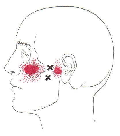
Trigger points in the lateral pterygoid

The lateral pterygoid muscle plays an important role in prober jaw function. It originates on the greater wing of the sphenoid bone and the lateral pterygoid plate, and inserts on the condyloid process of the mandible. It’s action is to pull the head of the mandibular condyle out of the mandibular fossa while opening the jaw. When trigger points develop they refer pain into the temporal mandibular joint and maxillary sinus. This referral is commonly mistaken for TM arthritis. In addition to the referral pain, trigger points in this muscle can also effect proper movement of the jaw.

FAI starts you on the road to failure.

Your hip socket or “acetabulum” is covered by smooth, glassy cartilage extending all the way to its outer rim. The term “Femoroacetabular impingement” (FAI) means that this rim of cartilage is being pinched when you move your hip into certain positions. Repetitive pinching results in irritation, tearing, or even detachment of this cartilage from your hip socket.

FAI occurs because of a mismatch between the head of your femur and the socket of your hip. The mismatch may be from an abnormally shaped hip bone (cam deformity), or having too much cartilage on the rim of your hip socket (pincer deformity). Most frequently, FAI results from a combination of both (combined or mixed deformity).

FAI is most common in young active people. Although the deformity may be present on both sides, symptoms are usually one-sided. The condition is equally common among men and women. Symptoms of FAI include a constant dull pain with periods of sharp pain, made worse by activity. Walking, pivoting, prolonged sitting, stair climbing, and impact activities like running or jumping can aggravate your symptoms. Snapping, locking, and clicking are common.

Our office will help direct a rehab program to maximize your chance of recovery. You should attempt to stay active, but avoid activities that aggravate symptoms. You may consider temporarily switching to low-impact activities, like stationary biking or water-walking. You should avoid motions that combine flexion and internal rotation, like- getting out of a car with one leg at a time, swimming the breaststroke, or performing squats. Patients with FAI should avoid excessive stretching, as this could aggravate the condition, but will likely benefit from strengthening exercises in the type of treatment provided in this office.

An Open Letter to our Medical friends.

462

In the past year, many trusted medical establishments including the FDA (1), CDC (2), Joint Commission (3,4), JAMA (5), and The American College of Physicians/ Annals of Internal Medicine (6) have encouraged medical providers to prescribe spinal manipulation as a first line treatment for acute, subacute, and chronic low back pain.

Most recently, The Lancet echoed that endorsement, and provided a unique perspective:

The reduced emphasis on pharmacological care recommends nonpharmacological care as the first treatment option and reserves pharmacological care for patients for whom nonpharmacological care has not worked. These guidelines endorse the use of exercise and a range of other non- pharmacological therapies, including massage, spinal manipulation, and acupuncture.

Gaps between evidence and practice exist, with limited use of recommended first- line treatments and inappropriately high use of imaging, rest, opioids, spinal injections, and surgery. Doing more of the same will not reduce back-related disability or its long-term consequences. The advances with the greatest potential are arguably those that align practice with the evidence. (7)

Unfortunately, personal experience skews our perception of each other’s merit, i.e., we primarily see each other’s failures since the successes don’t need to seek additional care. Regardless of our professional degree, we all have failed cases mixed into our many clinical successes. We must not lose sight of the evidence supporting each other’s overwhelming proven value for a given diagnosis. If we judge each other by our successes rather than our failures, we will work toward an integrated model where the patient wins. Together, we will help more patients than either working alone.

We are honored for the opportunity to co-manage your patients.


References
1. FDA Education Blueprint for Health Care Providers Involved in the Management or Support of Patients with Pain. May 2017. Accessed on May 12, 2017
2. Dowell D, Haegerich TM, Chou R. CDC Guideline for Prescribing Opioids for Chronic Pain- United States, 2016. MMWR Recomm Rep 2016;65(No. RR-1):1–49.
3. The Official Newsletter of The Joint Commission. Joint Commission Enhances Pain Assessment and Management Requirements for Accredited Hospitals. July 2017 Volume 37 Number 7. Ahead of print in
2018 Comprehensive Accreditation Manual for Hospitals.
4. Joint Commission Online. Revision to Pain Management Standards. http://www.jointcommission.org/assets/1/23/jconline_november_12_14.pdf
5. Paige NM, Miake-Lye IM, Booth MS, et al. Association of Spinal Manipulative Therapy With Clinical Benefit and Harm for Acute Low Back Pain; Systematic Review and Meta-analysis. JAMA. 2017;317(14):1451-1460.
6. Qaseem A, et al. for the Clinical Guidelines Committee of the American College of Physicians. Noninvasive Treatments for Acute, Subacute, and Chronic Low Back Pain: A Clinical Practice Guideline From the American College of Physicians. Ann Intern Med. 2017;166(7):514-530.
7. Foster, Nadine EBuchbinder, Rachelle et al. Prevention and treatment of low back pain: evidence, challenges, and promising directions. The Lancet, Published Online March 21, 2018 http://dx.doi.org/10.1016/ S0140-6736(18)30489-6

I’ve never baked anything in my life; how do I have a Baker’s Cyst?

Your body has strategically-placed, fluid-filled cushions called “bursa” around each joint. Bursa act to reduce friction where muscular tendons rub on bone. Your knee is surrounded by several bursa that share fluid with the inside of your knee joint and with each other. You can think of this like a chain of lakes connected by streams. If the inside of your knee joint or any of the bursa around your knee becomes inflamed, the entire system fills. When swelling exceeds each individual bursa’s capacity, this fluid creates a soft balloon-like swelling of the popliteal bursa behind your knee called a “Popliteal cyst”, or more commonly a “Baker’s cyst”.
This swelling is usually not from a problem with the popliteal bursa itself, but rather, inflammation somewhere else in the joint that has distended the popliteal bursa. This swelling is often the result of arthritis or damage to a ligament or cartilage within the knee.

A Baker’s cyst is often painful, and the discomfort may increase when you attempt to move your knee. Depending on the severity of your swelling, your symptoms may vary between a feeling of fullness and significant pain. It is not unusual for a Baker’s cyst to change in size from day to day, depending on the amount of swelling. Be sure to tell us if your pain progressively increases when you walk or if you notice warmth, redness or swelling in your calf, as these could be signs of a more significant problem.

Sometimes rest and elevation of your leg help to alleviate pain and swelling. Initially, you may need to avoid activities, like squatting, kneeling, heavy lifting, climbing stairs, or running. Wearing a brace on your knee may compress the cyst and cause an increase in your pain. Since Baker’s cysts are generally a problem that is secondary to another condition, treatment is most effective when directed at the underlying problem.

Spring Cleaning Tips

If you plan on doing a bit of spring cleaning soon, here is our friendly safety reminder: Don’t forget that some chores are a workout, so stretch accordingly before and after. Also, be mindful of your posture throughout your cleaning.

Keep a neutral spine, bend at your knees when lifting heavy items, and never twist your back at unhealthy angles.

Trigger points in the serratus anterior

The serratus anterior muscle is located along the sides of the ribs. It originates on the outer surface of the upper 8-9ribs, and inserts on the medial border of the scapula. This muscle acts on the scapula in several different ways. First it rotates the scapula to turn the glenoid fossa upward. It also protracted and elevates the scapula. And lastly it helps to prevent wining. This muscle is often shortened from prolonged sitting and work on a computer. Active trigger points in this muscle refer pain locally around the trigger point with spillover down the inside of the arm. Pain can also radiate into the inferior angle of the scapula.

There is nothing great about greater trochanteric pain syndrome. Nothing at all.

Your hip typically has about six small fluid-filled sacs called “bursa” that act as cushions between tendons and bone. One of the most common causes of hip pain is a condition called, “hip bursitis” which means that one or more of your bursas have become painfully inflamed. The broader (more accurate) diagnosis of “Greater trochanteric pain syndrome” (GTPS) describes an uncomfortable collection of problems affecting the outermost portion of your hip. GTPS can include swelling of one or more of the fibrous tendons that attach your muscles onto your hip – a condition called, “tendinitis.” In addition to bursitis & tendonitis, GTPS may originate from tightness in the muscle that travels over your hip en route to your knee- resulting in compression and irritation to your hip.


Greater trochanteric pain syndrome is most common in middle age to elderly adults and is 2-4 times more common in females. Sometimes the problem affects both hips at the same time. Approximately 1/3 of patients with GTPS have lower back pain. Patients who have arthritis in their hips and knees are more likely to suffer from ongoing complaints.

Your symptoms probably include a persistent pain on the outside of your hip, buttock, and upper thigh. Your discomfort may be aggravated by sitting with your leg crossed, arising from a seated position, prolonged standing, climbing stairs, and high-impact activities, like running. Sometimes patients find it difficult to sleep, since lying on the painful hip usually increases symptoms.

For adults, x-rays may or may not be needed to confirm the diagnosis, but children and adolescents usually require films to rule out more serious childhood diseases. Be sure to tell your doctor if you notice that you have a fever, leg numbness, pain radiating significantly beyond your knee, or pain in the front of your groin crease (the area where you leg meets your pelvis.)

Conservative treatment, like the type provided in this office, is successful in about 90% of cases. If you have acute pain, you may need to temporarily limit or discontinue activities that increase your discomfort. Using ice or ice massage at home may help. Some patients find temporarily relief by applying sports creams. Very commonly, patients with pain on the outside of their hip suffer from weakness in one of their buttock muscles, called the “gluteus medius.” When this muscle lacks strength, it is unable to protect your hip during normal activities, like walking. Research has shown that strengthening your hip has a dramatic effect on your progress.

Athletes should avoid running on a banked surface, like the crown of a road or indoor track. Be sure to reverse directions each mile if you run on a circular track. Avoid running on wet or icy surfaces, as this can cause increased tension in your hip. Runners with a “lazy” narrow-based running gait will benefit by increasing their step width to minimize stress on their hip. Cyclists need to make sure that their seat is not positioned too high. Overweight patients should consider weight reduction programs.

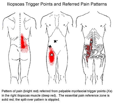
Trigger points in the iliopsoas muscle

This muscle originates on the bodies and disks of T12-L5 and the inner ilium. It inserts on the lesser trochanter of the femur. The psoas flexes the hip when the spine is fixed. When the leg is fixed it extends the lumbar spine increasing lumbar lordosis. This Muscle is often chronically shortened due to inactivity and sitting posture. When trigger points are present they will refer pain primarily to the lower lumbar area and the sacrum as well as into the anterior thigh. Trigger points in the iliopsoas muscle can mimic appendicitis.