How can I calm down my WAD symptoms?

Cervical Strain

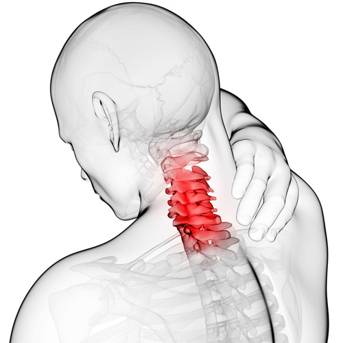
Whiplash is really a slang term for the rapid back and forth whipping of the head on the neck, usually associated with motor vehicle accidents. The title “Whiplash Associated Disorders”, or WAD, describes it best because it includes ALL of the MANY signs and symptoms of the disorder.

WAD basically comes in three sizes based on the degree of injury. A WAD I is present when there is pain but no physical examination findings; WAD II occurs when there are exam findings but no neurological loss (numbness or weakness); and WAD III includes loss of neurological function.  There is also a separate WAD level that includes fractures and dislocations (WAD IV).

There are many things that can be done by the patient to assist in the healing process for WAD. The first well-studied recommendation is to “continue with your usual activities.” Try to keep active and not change your routine. The good news is that WAD (especially types I and II) usually resolves without complication, and recovery is even more likely to occur if you don’t deviate much from your routine.

For those whose symptoms are more severe and/or not resolving, mobilization and manipulation of the neck and back are very effective treatment options. In addition to treatments you’d receive in a chiropractic office, there are MANY things you can do at home as “self-help strategies.” Some of these include (“PRICE”):

1)  PROTECT: Though it’s important to continue with your usual daily activities, this is dependent on both the degree of tissue injury and your pain tolerance. So do as many of your usual daily activities as possible, but AVOID those that result in a sharp, lancinating type of pain or those where recovery from the pain is delayed.  Therefore, this category may require modifying your ADLs (activities of daily living). A cervical collar (hard orsoft) should NOT to be used UNLESS you have an unstable injury (fracture or a grade III sprain).

2)  REST: Doing too much is like picking at a cut (which can delay healing) and doing too little can lead to a delayed healing response as well. Staying within reasonable pain boundaries is a good guide.

3) ICE > HEAT: Ice reduces swelling, and your doctor will typically recommend it over applying heat, especially on a recent injury. Heat draws fluids in, and while it may feel good, it can make your symptoms worse.

4)  COMPRESS: We can basically ignore this when referencing neck pain. This pertains better to wrapping an ankle, knee, wrist, or elbow with an elastic compression orthotic or brace.

5)  ELEVATE: This too is meant for the acute stages of an extremity injury like a foot or ankle.

Exercises unique for neck pain in the acute, subacute, and chronic stages of healing are perhaps the most important of the self-help approaches. In the ACUTE phase, try these…

1)  Range of Motion: Once again, stay within “reasonable pain boundaries” as you move your head forwards, backwards, side to side, and rotate left and right. These can be done either with or without LIGHT resistance applied using one or two fingers placed against your head. Limit the repetitions to three slow reps in each direction and emphasize the release of the movement.

2)  Chin/head Glides: Tuck in the chin (think of creating a double or triple chin) followed by poking the chin/head out.

In the SUBACUTE and CHRONIC phases of healing, the importance of strengthening the deep neck flexors cannot be over emphasized. Please refer to last week’s article for a description of this (see #3 of the 6 recommendations listed).

Our Exercise Of The Month: Dead Bug

Begin lying on your back with your right arm reaching overhead and your left leg flat on the table. Your right knee should be bent 90 degrees and your hip 45 degrees. Place your left wrist beneath your back to prevent your back from flattening against the ground. Slowly begin by raising your left knee and right arm at the same time until your hand touches your knee. Be sure not to lift your head or allow your spine to flatten against the floor. Return to the start position and repeat for three sets of 10 repetitions on each side, twice per day or as directed.

When Are My Shoes “Done”?

168

Running shoes need to be replaced every 250 miles. There are three basic options:


Motion Control Shoes – Designed for people with low or no arches, these shoes are for runners who strike the ground on the outer edge of their foot. Avoid overly stiff shoes as these decrease you perception of ground strike and lead to new injuries.


Stability or Neutral Shoes – Designed for people with normal or average arches and running mechanics. The shoe contains some cushioning to absorb shock and prevent injuries and some rigidity to avoid pronation.


Cushioned Shoes – Designed for people with high arched feet. Their footprint will typically leave a thin band along the foot’s edge. As they run weight is distributed from heel strike to the outer edge of the foot and small toes that bear the brunt of “lift off.” This shoe is more flexible and absorbs the shock created by the lack or rotation (under-pronation) created by their running style.

Golfer’s Elbow? Thats a thing?

Most of the muscles that flex your wrist are attached to a bony bump on the inside of your elbow called the “medial epicondyle.” Sometimes, through injury or overuse, the site where these muscles originate can become irritated or inflamed. This condition is called “medial epicondylitis”, or “golfer’s elbow.”

Although the condition is named “golfer’s elbow,” over 90% of those affected

are not even athletes, much less golfers. Nonetheless, the condition is more common in certain sports, especially golf, throwing, bowling, football, archery, and weight lifting. Occupations that require heavy gripping or repeated hand movements, like carpentry or typing, can predispose you to this condition. Smokers and people who are obese are more likely to experience this condition.

Medial epicondylitis is the most frequent cause of pain on the “inside” of your elbow but is 3-10 times less likely that its “outside” counterpart- lateral epicondylitis (i.e. “tennis elbow”). Medial epicondylitis is most common between the ages of 40 and 60. The condition strikes the dominant arm in over ¾ of cases. Your symptoms will likely include a dull aching pain over the bump on the inside of your elbow that becomes more intense with use. As the condition progresses, you may notice grip weakness or limitations when shaking hands, grasping objects, and opening jars. Be sure to tell your doctor if you notice numbness or tingling traveling toward your hand.

If left untreated, medial epicondylitis can last indefinitely. Studies show that up to 40% of untreated patients suffer prolonged discomfort, some as long as three years. Fortunately, conservative treatment like the type provided in our office is effective for relieving this condition. Our office may prescribe an elbow “counterforce brace” to help dampen stress to the irritated area. This type of brace should not be used in patients who have numbness or tingling radiating into their fingers. Some patients may benefit from a “wrist splint” that is used at night to allow your tendon to heal in a lengthened position. Using ice or ice massage over your elbow can help limit pain. Your home exercises will play an important role in your recovery.

Initially, you may need to avoid activities that cause an increase in pain, like gripping, lifting, golfing, throwing balls, or swinging a racquet. Be sure to warm up properly and stretch prior to any heavy physical activity. Smokers would benefit from quitting. Overweight patients should consider increasing their aerobic activity to shed excess pounds.

Entering & Exiting Your Vehicle

Ferrari_599_from_Lusso

Entering and exiting your vehicle is a potential risky activity for low back pain sufferers. Follow these tips to limit problems:


To enter the vehicle, open the door and stand with your back to the seat, legs close to the side of the vehicle. For larger vehicles, you may wish to begin by standing on the running board. Place your hands on the door and door frame to keep your movements slow and controlled then slowly lower your body into the vehicle.

Tuck your head into the vehicle. Keep your knees close to each other, as though they have been taped together, brace your abdomen as though you are about to be punched in the stomach and pivot your body as a whole without twisting or bending at the waist. You may grasp the steering wheel with your right hand to help you pivot.

Use a lumbar roll or other support to help maintain good posture. Position the roll slightly above your belt to support the “small of your back”. Adjust your seat so that your knees are slightly lower than your hips. Try to avoid prolonged car rides- take frequent breaks.

Before exiting, create adequate space by pushing your vehicle seat back as far as possible and move the steering wheel up and out of the way. To exit, first scoot slightly to the door side edge of your seat, then keep your knees together and pivot with the same cautions that you used to enter the vehicle. When your feet are shoulder width apart and firmly on the ground or running board, grasp the door and door frame, lean forward, but be sure not to bend your back, as you tighten your abdominal muscles. Slowly thrust your hips forward to stand up.

Workers compensation cases highlight the value of chiropractic care

160801-F-DB969-034

A newly published analysis of 5511 workers compensation cases highlights the value of chiropractic care: “those injured workers who first saw a chiropractor experienced shorter first episodes of compensation (vs. physical therapy alone)”.

If you or someone you know has suffered a job-related injury, call us today. Our goal is to provide the most effective care that gets you back to work ASAP!

https://www.ncbi.nlm.nih.gov/pubmed/27638518

Do you have Carpal Tunnel Syndrome?

Carpal Tunnel Syndrome is VERY common. If fact, it affects 3-6% of the general population, and up to 50% of those afflicted have it in both hands! So what makes it so common? Here are several reasons:

1) ANATOMY: The wrist is made up of eight small “carpal” bones that allow the wrist to bend and twist in many different positions. If you were to look at the wrist “head on,” you’d notice it is shaped like a tunnel where the eight little carpal bones make up the “roof” and the transverse carpal ligament is the “floor.” You’ll also notice nine tendons and their sheaths that pass through the carpal tunnel and attach the forearm muscles to the fingers on the palm side of the arm, blood vessels, and the median nerve. When there is less available space within the carpal tunnel (due to inflamed tendons, for example), the added pressure can compress the median nerve and result in numbness, tingling, and weakness, mostly into the second to fourth fingers.

2) JOBS: People with occupations that require fast, repetitive movements and/or a firm grip with little rest have a higher likelihood of developing CTS.

3) GENDER: Women have a greater risk of developing CTS than men due to smaller wrists and hormonal changes that may result in swelling in the wrist.

4) AGE: Individuals over age 50 have a greater risk of developing CTS, especially females.

5) GENERAL HEALTH ISSUES: There are a host of conditions that can cause and/or aggravate CTS. Some of these include rheumatoid arthritis, diabetes, osteoarthritis, low thyroid function, and space-occupying issues like a ganglionic cyst inside the tunnel. Certain medications, such as birth control pills, and some forms of chemotherapy can also cause CTS. A pinched nerve in the neck or shoulder can alter median nerve function, which can exacerbate CTS symptoms.

6) TRAUMA: A fall onto an outstretched arm can sprain and/or fracture the wrist. If a fracture occurs, the onset of CTS can be very rapid, and quick realignment of the fracture is very important.

Chiropractic care offers a non-surgical solution to CTS, which includes splinting (primarily at night and when driving but usually less often during the day as we tend to “fight” against the splint), anti-inflammatory measures (ice, nutrients such as ginger, turmeric, bioflavonoids), and manual therapies—manipulation of the fingers, hand, wrist, elbow, shoulder, and neck (as indicated) and muscle release techniques to the forearm are very helpful. There are also effective exercises that patients can do at home that include joint range of motion and muscle stretching techniques.


Shrug

Trigger points in the trapezius muscle.

The trapezius muscle is a large diamond shaped muscle in you mid/upper back and neck. This muscle is one of the most common sites where trigger points can form. This muscle originates on the Nuchal ligament and the spinous processes of C6-T12. It inserts on the spine of the scapula, the acromion process, and the distal clavicle. The upper fibres elevate the shoulder and rotate the glenoid fossa (shoulder socket) upward. The lower fibres assist this motion as well as help depress the shoulder. The middle fibres of this muscle strongly adduct the scapula. This muscle is susceptible to postural overload such as sitting at desk all day. Trigger points on the lateral upper edge refer into the lateral neck and temples, causing “tension neck ache”as well as headache pain.Trigger points in the middle and lower fibres refer pain into the posterior neck and shoulder.

Our Condition of the Month: ITB Syndrome

Iliotibial Band Syndrome

Your iliotibial band is a fibrous band of tissue running from the crest of your hip, down to your knee. A muscle near the crest of your hip called the tensor fascia lata, attaches to this band to help control movements of your leg. Your iliotibial band passes over the point of your hip and over another bony bump on the outside of your knee called the lateral epicondyle. When the band is too tight, it can become painfully inflamed as it repeatedly rubs over the top of either of these bony projections. Irritation at the bony bump near the knee is called iliotibial band friction syndrome.

The condition usually presents as pain on the outside of your knee that becomes worse with repetitive knee flexion or extension. This condition is the leading cause of lateral knee pain in runners, especially slower “joggers.” The pain usually develops 10-15 minutes into the workout. You may sometimes feel or hear a click during movement. Symptoms generally ease at rest. Running on slick “wintry” surfaces may aggravate the condition. Excessively worn running shoes may be a culprit.

This problem is commonly caused by weakness in your gluteal muscles. When these muscles are weak, the muscle that attaches to your iliotibial band must contract harder to stabilize your hip. Having one leg longer than another is a known aggravating factor.

Runners should minimize downhill running and avoid running on a banked surface like the crown of a road or indoor track, as well as wet or icy surfaces. Runners should reverse directions on a circular track at least each mile. Bicyclists may need to adjust seat height and avoid “toe in” pedal positions. Avoid using stair climbers or performing squats and dead lifts. Sports cream and home ice massage may provide some relief of symptoms.