Where is my Low Back Pain coming from?

​

Most of us have suffered from back pain at one time or another. It often occurs after over-doing a physical task, like fall yard work, winter snow shovelling, working on the car, cleaning the house, and so on. But there are times when identifying the cause of back pain can be difficult or impossible. Let’s take a deeper look at where back pain can come from…
Though activity-related back pain is common, many times a direct link to over-use is not clear. Micro-traumatic events can accumulate and become painful when a certain threshold is exceeded. (Think of the old adage “The straw that broke the camel’s back.”)
There are other less well-identified causes of back pain. One is called referred pain. This can be caused by an irritated joint or soft tissue not necessarily located in the immediate area of the perceived pain. For example, pain in the leg can result from an injured facet joint, sacroiliac joint, and/or a disk tear (without nerve root pinch). This is called “sclertogenous pain.”
Internal organs can also cause back pain. This is called a “viscerosomatic response” (VSR). A classic example of this is when the right shoulder blade seems to be the source of pain when the gall bladder is inflamed. This pain can be located at or below the scapula next to the spine and the muscles in the area are in spasm and sensitive or painful to the touch. Also, VSR is often not worsened or changed by bending in different directions (unlike musculoskeletal / MSK pain). Without further testing, it’s easy to confuse this with a MSK or a “typical” back ache. Ultimately, a final diagnosis may require an abdominal ultrasound (CT, MRI scan, and other diagnostics are less frequently used).
Visceral pathology in the back pain patient presenting to chiropractors is reportedly rare, and according to one survey, only 5.3% of patients present with non-musculoskeletal complaints. Other common VSR pain patterns are as follows: Heart – left chest to left arm, mid-upper back, left jaw; Liver – right upper shoulder (front and back), right middle to low back, and just below the sternum; Appendix – right lower abdomen (may start as stomach pain); Small intestine – either side of the umbilicus and/or between it and the breast bone; Kidney – small of the back, upper tailbone, and/or groin area; Bladder – just above the pubic bone and/or bilateral buttocks; Ovaries – groin and/or umbilical area; and Colon – mid-abdominal and/or lower quadrants.
Another challenge to diagnosis is cancer in the spine, which can be primary or metastatic (from a different location). Thankfully, this is very rare. A history of unexplained weight loss, a past history of cancer, over age 50, nighttime sleep interruptions, and no response to usual back care may lead a doctor to recommend tests to determine if cancer is present in the spine.
Bottom line: When patients present with back pain, chiropractors have been trained to look for these less common but important causes of back pain. They get “suspicious” when the “usual” orthopedic tests do not convey the usual responses seen with mechanical back pain. In these cases, they work with primary care doctors to coordinate care to obtain prompt diagnostic testing and treatment.
We realize you have a choice in whom you consider for your health care provision and we sincerely appreciate your trust in choosing our service for those needs.  If you, a friend, or family member requires care for back pain, we would be honored to render our services.

361

Exercise of the Month!

One of our favourites in the office; Thoracic Rotation.

An easy exercise for your upper and mid-back. No equipment required! – (Post link to article and video on your blog or post video directly on your social media account.)”

  1. Begin on your hands and knees and shift back so that your buttock is on your heels.
  2. Place one hand behind your neck with your elbow pointing downward.
  3. Rotate your trunk to move your elbow towards your opposite knee.
  4. Rotate back, raising your elbow toward the ceiling.
  5. Repeat three sets of 10 repetitions on each side twice per day or as directed.

Pregnancy and Low Back Pain?

40​

Did you know that 50-72% of women have low back pain (LBP) and/or pelvic pain during their pregnancy but only 32% do something about it? Let’s look closer!
Pregnancy-related low back pain (PLBP) can be a highly debilitating syndrome that accounts for the most common cause of sick leave for pregnant women. In 2004, Americans spent $26.1 billion dollars in an effort to find relief from back pain during pregnancy. Statistics show one out of ten women will experience daily DISABLING LBP for at least two years following delivery.
Because of the limited number of treatment options available for the pregnant woman due to mother and fetus safety, and given the high propensity of potentially disabling PLBP that can significantly limit function and quality of life, chiropractic care seems to be a natural choice for this patient population! Obviously, pharmaceuticals and surgery are NOT appropriate options for the expectant mother, even during the post-partum breast-feeding time period. Chiropractic offers a non-invasive and safe approach to managing lumbopelvic pain that uses many different approaches.
In a 2009 research paper, 78 women participated in a study that investigated disability, pain intensity, and percent improvement after receiving chiropractic care to treat pregnancy-related PLBP. Here, 73% reported their improvement as either “excellent” or “good.” For disability and pain, 51% and 67% (respectively) experienced clinically significant improvement! Researchers followed up with them eleven months later and found 85.5% reported their improvement as either “excellent” or “good!” For disability and pain, 73% and 82% (respectively) experienced clinically significant improvement!
So, what’s causing LBP in pregnant women? Because of the biomechanical changes that occur in the low back and pelvis over a relatively short amount of time during pregnancy, especially in the second and third trimesters, common pain generators include (but are not limited to): the sacroiliac joint, facet joints, shock-absorbing disks, and the many connecting muscles (strains) and ligaments (sprains). During the later stages of pregnancy, the hormone Relaxin prepares the pelvis for delivery by widening the pelvic girdle, which can also be problematic.
Treatment options within chiropractic often include spinal manipulation, lumbopelvic exercises, patient education, posture correction, massage, an SI belt, soft tissue mobilization, and more. Exercises that target the transverse abdominus, multifidus, and pelvic floor muscles help to stabilize the lumbopelvic region. The American College of Obstetricians & Gynaecologists recommends exercise at least three times a week during pregnancy, and studies report NO obstetric complications (pre-term labor, premature ruptured membranes, or changes to maternal or neonatal weight) with exercise.
So, the answer is clear! When PLBP strikes, seek chiropractic care to safely and effectively manage the pain and disability and so you can ENJOY YOUR PREGNANCY!!!
We realize you have a choice in whom you consider for your health care provision and we sincerely appreciate your trust in choosing our service for those needs.  If you, a friend, or family member requires care for back pain, we would be honoured to render our services.

Condition Of The Month: Thoracic Joint Restriction

T Spine Joint

Your spine is made up of 24 bones stacked on top of each other with a soft “disc” between each segment to allow for flexibility. Normally, each joint in your spine should move freely and independently. When one or more of your spinal vertebra is slightly misaligned and restricted, we call this condition a “spinal segmental joint restriction”. A “thoracic joint restriction” means that this misalignment or restriction is located in your upper or mid-back region.

To help visualize this, imagine a normal spine functioning like a big spring moving freely in every direction. A spine with a joint restriction is like having a section of that spring welded together. The spring may still move as a whole, but a portion of it is no longer functioning.

Joint restrictions can develop in many ways. Sometimes they are brought on by an accident or an injury. Other times, they develop from repetitive strains or poor posture. Being overweight, smoking, strenuous work, and emotional stress can make you more susceptible to problems.

Restricted joints give rise to a self-perpetuating cycle of discomfort. Joint restriction causes swelling and inflammation, which triggers muscular guarding leading to more restriction. Since your spine functions as a unit, rather than as isolated pieces, a joint restriction in one area of your spine often causes “compensatory” problems in another. Think of this as a rowboat with multiple oarsmen on each side. When one rower quits, the others are placed under additional stress and can become overworked.

Joint restrictions most commonly cause local tenderness and discomfort. You may notice that your range of motion is limited. Movement may increase your discomfort. Pain from a restricted joint often trickles around your rib cage or up & down your spine. Be sure to tell your chiropractor if your symptoms include any chest pain, shortness of breath, unusual cough, indigestion or flu- like complaints.

Long-standing restrictions are thought to result in arthritis – much like the way a slightly misaligned wheel on your car causes premature wearing of your tire.

You should recognize that your problem is common and generally treatable. Chiropractic care has been shown to be the safest and most effective treatment for joint restrictions. Our office offers several tools to help ease your pain. To speed your recovery, you should avoid activities that increase your pain. Be sure to take frequent breaks from sedentary activity. Yoga has been shown to help back pain sufferers so consider joining a class or picking up a DVD.

Chiropractic as THE alternative to Opioids.

In 2015, two million Americans had a substance abuse disorder involving prescription pain relievers; with more than 20,000 overdose deaths related to these drugs. In the past decade, death rates and substance use rates quadrupled in parallel to sales of prescription pain relievers.

Chronic LBP is a primary generator for opioid prescriptions. This year, both JAMA and Annals of Internal Medicine have published and supported new clinical practice guidelines that recommend prescribing spinal manipulation over medication for LBP patients.

“For acute, subacute, or chronic low back pain, physicians and patients initially utilize spinal manipulation and delay pharmacologic management.”

Research shows that low back pain patients who undergo chiropractic care have improved outcomes with lower rates of opioid use, surgery, and overall healthcare costs.

Not surprisingly, various governing bodies, including the FDA, CDC, and 37 State Attorney General’s, have concurred that physicians and healthcare decision makers should consider non- pharmacologic therapy for LBP patients. In fact, the 2018 Joint Commission guidelines mandate that hospitals include conservative options for chronic musculoskeletal pain management, specifically naming chiropractic as a potential option.

Chiropractic care is not a replacement for traditional medical treatment of LBP, rather a complementary tool to integrate within your current management paradigm. We hope that you will continue to consider our office for those cases that may be favorably served by conservative manual therapy. We are grateful for your confidence and will work hard to maintain your trust.

Do I Need an MRI?

Low back pain is a very common complaint. In fact, it’s the #1 reason for doctor visits in the United States! The economic burden of LBP on the working class is astronomical. Most people can’t afford to be off work for one day, much less a week, month, or more! Because of the popularity of hospital-based TV dramas over the past two decades, many people think getting an MRI of their back can help their doctor fix their lower back problem. Is this a good idea? Let’s take a look!
Patients will often bring in a CD that has an MRI of their lower back to a doctor of chiropractic and ask the ultimate question, “….can you fix me?” Or, worse, “…I think I need surgery.” Sure, it’s quite amazing how an MRI can “slice” through the spine and show bone, soft tissues, disks, muscles, nerves, the spinal cord, and more! Since the low back bears approximately 2/3 of our body’s weight, you can frequently find MANY ABNORMALITIES in a person over 40-50 years old. In fact, it would be quite odd NOT to see things like disk degeneration, disk bulges, joint arthritis, spur formation, etc.!
Hence, the “downside” of having ALL this information is the struggle to determine which finding on the MRI has clinical significance. In other words, where is the LBP coming from? Is it that degenerative disk, bulged disk, herniated disk, or the narrowed canal where the nerve travels? Interestingly, in a recent review of more than 3,200 cases of acute low back pain, those who had an MRI scan performed earlier in their care had a WORSE outcome, more surgery, and higher costs compared with those who didn’t succumb to the temptation of requesting an MRI!
This is not to say MRI, CT scans, and x-rays are not important, as they effectively show conditions like subtle fractures and dangerous conditions like cancer. But for LBP, MRI is often misleading. This is because the primary cause of LBP is “functional” NOT “structural,” so it’s EASY to get railroaded into thinking whatever shows up on that MRI has to be the problem.
Here is how we know this, when we take 1,000 people WITHOUT low back pain between ages 30 and 60 (male or female) and perform an MRI on their lower back, we will find up to 53% will have PAINLESS disk bulges in one or more lumbar disks. Moreover, we will find up to 30% will have partial disk herniations, and up to 18% will have an extruded disk (one that has herniated ALL the way out). Yet, these people are PAIN FREE and never knew they had disk “derangement” (since they have no LBP). When combining all of these possible disk problems together, several studies report that between 57% and 64% of the general population has some type of disk problem without ANY BACK PAIN!
Hence, when a patient with a simple sprain/strain and localized LBP presents with an MRI showing a disk problem, it usually ONLY CONFUSES the patient (and frequently the doctor), as that disk problem is usually not the problem causing the pain!  So DON’T have an MRI UNLESS a surgical treatment decision depends on its findings. That is weakness, numbness, and non-resolving LBP in spite of 4-6 weeks of non-surgical care or unless there is weakness in bowel or bladder control. Remember, the majority of back pain sufferers DO NOT need surgery!
We realize you have a choice in whom you consider for your health care provision and we sincerely appreciate your trust in choosing our service for those needs.  If you, a friend, or family member requires care for back pain, we would be honored to render our services.

PFPS Cont. You want details?

Screen Shot 2017-10-31 at 11.15.32 AM.png

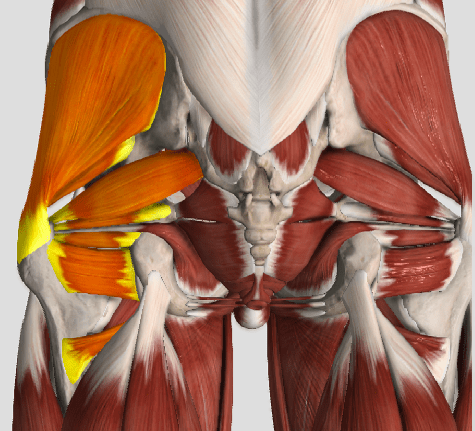
The muscles of the hip provide not only local stability, but also play an important role in spinal and lower extremity functional alignment. (1-4) While weakness in some hip muscles (hip extensors and knee extensors) is well tolerated, weakness or imbalance in others can have a profound effect on gait and biomechanical function throughout the lower half of the body. (5) Weakness of the hip abductors, particularly those that assist with external rotation, has the most significant impact on hip and lower extremity stability. (5,6)

Hip Abduction.gif

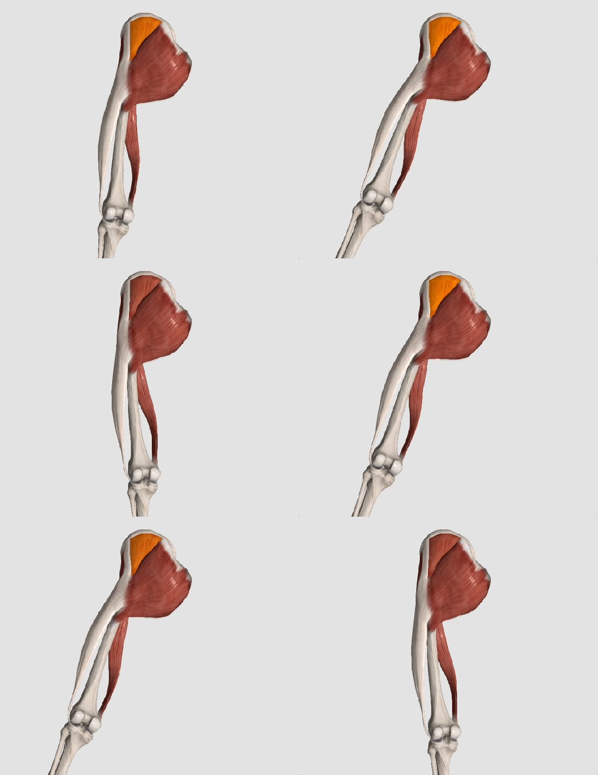
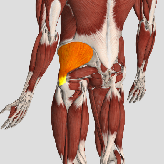
The gluteus medius is the principal hip abductor. When the hip is flexed, the muscle also assists the six deep hip external rotators (piriformis, gemelli, obturators, and quadratus femoris). The gluteus medius originates on the ilium just inferior to the iliac crest and inserts on the lateral and superior aspects of the greater trochanter. While the principal declared action of the gluteus medius is hip abduction, clinicians will appreciate its more valuable contribution as a dynamic stabilizer of the hip and pelvis- particularly during single leg stance activities like walking, running, and squatting. The gluteus medius contributes approximately 70% of the abduction force required to maintain pelvic leveling during single leg stance. The remainder comes predominantly from 2 muscles that insert onto the iliotibial band: the tensor fascia lata and upper gluteus maximus.  Hip abductor strength is the single greatest contributor to lower extremity frontal plain alignment during activity. (6)

Screen Shot 2017-10-31 at 11.12.00 AM

Incompetent hip abductors and/or external rotators allows for excessive adduction and internal rotation of the thigh during single leg stance activities. This leads to a cascade of biomechanical problems, including pelvic drop, excessive hip adduction, excessive femoral internal rotation, valgus knee stress, and internal tibial rotation. (1,7-12)


References
1. Szu-Ping Lee, Powers C. Description of a Weight-Bearing Method to Assess Hip Abductor and External Rotator Muscle Performance. JOSPT. Volume 43, Issue 6
2. Crossley KM, Zhang WJ, Schache AG, Bryant A, Cowan SM. Performance on the single-leg squat task indicates hip abductor muscle function. Am J Sports Med. 2011;39:866-873.
3. Presswood L, Cronin J, Keogh JWL, Whatman C. Gluteus medius: applied anatomy, dysfunction, assessment, and progressive strengthening. Strength Cond J. 2008;30:41-53.
4. Sled EA, Khoja L, Deluzio KJ, Olney SJ, Culham EG. Effect of a home program of hip abductor ex- ercises on knee joint loading, strength, function, and pain in people with knee osteoarthritis: a clinical trial. Phys Ther. 2010;90:895-904.
5. van der Krogt MM, Delp SL, Schwartz MH How robust is human gait to muscle weakness? Gait Posture. 2012 Feb 29.
6. Laurie Stickler, Margaret Finley, Heather Gulgin Relationship between hip and core strength and frontal plane alignment during a single leg squat Physical Therapy in Sport Available online 2 June 2014
7. Ireland ML, Willson JD, Ballantyne BT, Davis
IM. Hip strength in females with and without patellofemoral pain. J Orthop Sports Phys Ther. 2003;33:671-676.
8. Noehren B, Davis I, Hamill J. ASB clinical biome- chanics award winner 2006: prospective study of the biomechanical factors associated with iliotib- ial band syndrome. Clin Biomech (Bristol, Avon). 2007;22:951-956.
9. Powers CM. The influence of abnormal hip me- chanics on knee injury: a biomechanical perspec- tive. J Orthop Sports Phys Ther. 2010;40:42-51.
10. Powers CM. The influence of altered lower- extremity kinematics on patellofemoral joint dysfunction: a theoretical perspective. J Orthop Sports Phys Ther. 2003;33:639-646.
11. Sigward SM, Powers CM. Loading characteristics of females exhibiting excessive valgus moments during cutting. Clin Biomech (Bristol, Avon). 2007;22:827-833
12. Souza RB, Powers CM. Differences in hip kine- matics, muscle strength, and muscle activation between subjects with and without patellofemo- ral pain. J Orthop Sports Phys Ther. 2009;39:12- 19.