5 Tips For Gym Newbies

3-Ways-to-Protect-Your-Joints-and-Avoid-Injuries-269x169

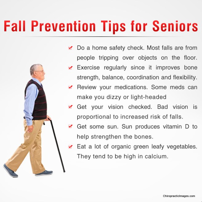
Michelle Blood has a great piece for new gym members over at LifeZette.com Check out the link at the end of the page for the whole article. Getting started can be a daunting task for many; these 5 tips will help you stay motivated, safe, on task and get you to your goals.

1.) Get checked. Before you set foot into the gym to begin any workout program, it is important you get clearance from your doctor. It is easy to overlook this step in the excitement over the idea of committing yourself to getting fit. However, failure to do so can be very costly. For the following groups of people, it is extremely important to hit the doctor’s office before you hit the gym:

  • You haven’t had a physical in the past year.
  • You’re planning a significant increase in the intensity of exercise.
  • You’re undertaking a new form of exercise.
  • You have a physical condition that may be exacerbated by exercise.
  • You have concerns about your physical capability for exercise.

2.) Get comfortable. Gyms can feel intimidating when you’re new because the layout and procedures at your gym are unfamiliar, as are the pieces of equipment and the group-exercise formats. The best way to combat these sorts of concerns is to take some time to tour the gym, to observe different group-based classes, and to receive some basic instruction on use of various pieces of equipment.

Consider approaching instructors and trainers. Most are friendly and personable, and more than willing to answer any questions you have about classes or equipment. Your fellow gym members can be an indispensable source of information as well. Ask them about their experiences in a class that interests you.

By getting as much information as you can prior to joining a class, you’ll feel more comfortable participating when you make the leap and sign up for your first group class.

3.) Get equipped. Fortunately, you don’t need to break the bank to gather up a few essentials you’ll want to have when you begin your gym-based fitness journey. Though you’re not going to need an entire new wardrobe, it would be a good idea to pick up a couple of sweat-wicking items (e.g., shorts, T-shirts) and an appropriate pair of shoes.

Other items on your shopping list might include: a filtered, refillable water bottle; showering necessities; a padlock (if your gym provides lockers); and some pre and post-workout healthy snacks. Store the essentials in a sturdy bag to throw in your trunk so you’ll always be ready to hit the gym for a quick workout whenever the mood may strike you.

4.) Get a partner. Building some accountability in to your new gym routine is a great way to bolster your chances of success. Bringing a friend or family member along to the gym has a number of advantages. When you commit to another person, you give yourself the gift of subtle, positive pressure to stick to your goals.

If you’re waffling about attending class on a given day, the fact that your partner will be there waiting for you can provide that extra bit of encouragement you need to power through your reluctance and show up for class anyway.

In addition, developing a fun competition with your partner can have some spectacular results. Science has shown us, time and again, that people lose more weight when they are involved with a team or are in a competition. Be sure to keep it positive, and you can spur one another to successes neither partner might have hoped to achieve on his or her own.

5.) Get pumped. You can beat the “I just don’t feel motivated” monster in a number of ways. Finding an effective strategy for motivating yourself is somewhat a matter of trial and error. Try some of the following to discover what works best for you.

  • Create a personalized music playlist of songs that get you moving.
  • Read books and articles about people who have achieved what you hope to achieve.
  • Set up a system of rewards for yourself for meeting small goals.
  • Spend time with people who inspire you.
  • Keep a journal of your feelings before and after working out — review it when you feel tempted to skip.

Regardless of your level of experience — you can confidently succeed at the gym.


http://www.lifezette.com/healthzette/success-at-the-gym-five-top-tips-for-exercise-newbies/

Chiro & Concussions

head-shot2

Whiplash Associated Disorders (WAD) is the appropriate terminology to use when addressing the myriad of symptoms that can occur as a result of a motor vehicle collision (MVC). In a recent publication in The Physician and Sports Medicine (Volume 43, Issue 3, 2015; 7/3/15 online:1-11), the article “The role of the cervical spine in post-concussive syndrome” takes a look at the neck when it’s injured in a car accident and how this relates to concussion.

It’s estimated about 3.8 million concussion injuries, also referred to as “mild traumatic brain injury” (mTBI), occur each year in the United States. Ironically, it’s one of the least understood injuries in the sports medicine and neuroscience communities. The GOOD NEWS is that concussion symptoms resolve within 7-10 days in the majority of cases; unfortunately, this isn’t the case with 10-15% of patients. Symptoms can last weeks, months, or even years in this group for which the term “post-concussive syndrome” (PCS) is used (defined as three or more symptoms lasting for four weeks as defined by the ICD-10) or three months following a minor head injury (as defined by the Diagnostic and Statistical Manual of Mental Disorders).

There have been significant advances in understanding what takes place in the acute phase of mTBI, but unfortunately, there is no clear physiological explanation for the chronic phase. Studies show the range of force to the head needed to cause concussion is between 60-160g (“g” = gravity) with 96.1g representing the highest predictive value in a football injury, whereas as little as 4.5g of neck acceleration can cause mild strain injury to the neck. In spite of this difference, the signs and symptoms reported by those injured in low-speed MVCs vs. football collisions are strikingly similar!

Research shows if an individual sustains an injury where the head is accelerated between 60-160g, it is HIGHLY likely that the tissues of the cervical spine (neck) have also reached their injury threshold of 4.5g. In a study that looked at hockey players, those who sustained a concussion also had WAD / neck injuries indicating that these injuries occur concurrently. Injuries to the neck in WAD include the same symptoms that occur in concussion including headache, dizziness/balance loss, nausea, visual and auditory problems, and cognitive dysfunction, just to name a few.

The paper concludes with five cases of PCS that responded well to a combination of active exercise/rehabilitation AND passive manual therapy (cervical spine manipulation). The favourable outcome supports the concept that the neck injury portion of WAD is a very important aspect to consider when treating patients with PCS!

This “link” between neck injury and concussion explains why chiropractic care is essential in the treatment of the concussion patient! This is especially true when the symptoms of concussion persist longer than one month!

We realize you have a choice in whom you consider for your health care provision and we sincerely appreciate your trust in choosing our service for those needs.  If you, a friend, or family member requires care for Whiplash, we would be honoured to render our services.

PFPS Cont. You want details?

Screen Shot 2017-10-31 at 11.15.32 AM.png

The muscles of the hip provide not only local stability, but also play an important role in spinal and lower extremity functional alignment. (1-4) While weakness in some hip muscles (hip extensors and knee extensors) is well tolerated, weakness or imbalance in others can have a profound effect on gait and biomechanical function throughout the lower half of the body. (5) Weakness of the hip abductors, particularly those that assist with external rotation, has the most significant impact on hip and lower extremity stability. (5,6)

Hip Abduction.gif

The gluteus medius is the principal hip abductor. When the hip is flexed, the muscle also assists the six deep hip external rotators (piriformis, gemelli, obturators, and quadratus femoris). The gluteus medius originates on the ilium just inferior to the iliac crest and inserts on the lateral and superior aspects of the greater trochanter. While the principal declared action of the gluteus medius is hip abduction, clinicians will appreciate its more valuable contribution as a dynamic stabilizer of the hip and pelvis- particularly during single leg stance activities like walking, running, and squatting. The gluteus medius contributes approximately 70% of the abduction force required to maintain pelvic leveling during single leg stance. The remainder comes predominantly from 2 muscles that insert onto the iliotibial band: the tensor fascia lata and upper gluteus maximus.  Hip abductor strength is the single greatest contributor to lower extremity frontal plain alignment during activity. (6)

Screen Shot 2017-10-31 at 11.12.00 AM

Incompetent hip abductors and/or external rotators allows for excessive adduction and internal rotation of the thigh during single leg stance activities. This leads to a cascade of biomechanical problems, including pelvic drop, excessive hip adduction, excessive femoral internal rotation, valgus knee stress, and internal tibial rotation. (1,7-12)


References
1. Szu-Ping Lee, Powers C. Description of a Weight-Bearing Method to Assess Hip Abductor and External Rotator Muscle Performance. JOSPT. Volume 43, Issue 6
2. Crossley KM, Zhang WJ, Schache AG, Bryant A, Cowan SM. Performance on the single-leg squat task indicates hip abductor muscle function. Am J Sports Med. 2011;39:866-873.
3. Presswood L, Cronin J, Keogh JWL, Whatman C. Gluteus medius: applied anatomy, dysfunction, assessment, and progressive strengthening. Strength Cond J. 2008;30:41-53.
4. Sled EA, Khoja L, Deluzio KJ, Olney SJ, Culham EG. Effect of a home program of hip abductor ex- ercises on knee joint loading, strength, function, and pain in people with knee osteoarthritis: a clinical trial. Phys Ther. 2010;90:895-904.
5. van der Krogt MM, Delp SL, Schwartz MH How robust is human gait to muscle weakness? Gait Posture. 2012 Feb 29.
6. Laurie Stickler, Margaret Finley, Heather Gulgin Relationship between hip and core strength and frontal plane alignment during a single leg squat Physical Therapy in Sport Available online 2 June 2014
7. Ireland ML, Willson JD, Ballantyne BT, Davis
IM. Hip strength in females with and without patellofemoral pain. J Orthop Sports Phys Ther. 2003;33:671-676.
8. Noehren B, Davis I, Hamill J. ASB clinical biome- chanics award winner 2006: prospective study of the biomechanical factors associated with iliotib- ial band syndrome. Clin Biomech (Bristol, Avon). 2007;22:951-956.
9. Powers CM. The influence of abnormal hip me- chanics on knee injury: a biomechanical perspec- tive. J Orthop Sports Phys Ther. 2010;40:42-51.
10. Powers CM. The influence of altered lower- extremity kinematics on patellofemoral joint dysfunction: a theoretical perspective. J Orthop Sports Phys Ther. 2003;33:639-646.
11. Sigward SM, Powers CM. Loading characteristics of females exhibiting excessive valgus moments during cutting. Clin Biomech (Bristol, Avon). 2007;22:827-833
12. Souza RB, Powers CM. Differences in hip kine- matics, muscle strength, and muscle activation between subjects with and without patellofemo- ral pain. J Orthop Sports Phys Ther. 2009;39:12- 19.

Patellofemoral Pain Syndrome

knock knee

Patellofemoral pain syndrome (PFPS), also called “Runners Knee”, describes the symptom complex of knee discomfort, swelling, or crepitus that results from excessive or imbalanced forces acting on the joint. It is the most common cause of knee pain in the general population, affecting an estimated 25% of adults.

PFPS is most commonly related to lateral tracking of the patella. The patella has a natural tendency to migrate laterally due to the pull of the quadriceps and the slight natural valgus of the lower extremity. A new study in the Journal of Sports Medicine (1) provides additional confirmation that when managing patellofemoral pain syndrome, clinicians must address two critical yet often overlooked issues.

This study concludes that PFPS and dynamic knee valgus do not arise primarily from knee dysfunction, rather from hip abductor/ external rotator weakness and/or foot hyperpronation.

“The most effective intervention programs included exercises targeting the hip external rotator and abductor muscles and knee extensor muscles.” and “PFPS patients with foot abnormalities, such as those with increased rearfoot eversion or pes pronatus, may benefit the most from foot orthotics.”

Since gluteus medius and VMO weakness are key factors in the development of PFPS, strengthening exercises that target those muscles prove most effective. Stabilization exercises may include pillow push (push the back of your knee into a pillow for 5-6 seconds), supine heel slide, terminal knee (short-arc) extension, clam, glut bridge, semi-stiff deadlift, posterior lunge, and monster walk.

Myofascial release and stretching should be directed at hypertonic muscles, including the TFL, gastroc, soleus, hamstring, piriformis, hip rotators, and psoas. Myofascial release or IASTM may be appropriate for tightness in the iliotibial band, vastus lateralis, posterior hip capsule, and lateral knee retinaculum.

Manipulation may be necessary for restrictions in the lumbosacral and lower extremity joints. Hypermobility is common in the ipsilateral SI joint with restrictions present contralaterally. Evidence has shown that patellar tracking braces, i.e. BioSkin® or PatellaPro®, may lead to better outcomes.

Lifestyle modification may be necessary to reduce pain-provoking endeavors, especially running, jumping and other activities that induce a valgus stress. Athletes should avoid allowing their knee to cross in front of their toes while squatting. Arch supports or custom orthotics may be necessary to correct hyperpronation. Runners should avoid cross-over gaits and change shoes every 250 to 500 miles.

.

.

.

.

.

.

.

#pain

#kneepain

#chiropractic

#fitness

#sports

#wellness

#fitness

#healthyliving

#healthychoices

#Winnipeg

References

1. Petersen W, Rembitzki I, Liebau C. Patellofemoral pain in athletes. Open Access Journal of Sports Medicine. 2017;8:143-15

Exercise and Low Back Pain

Low back pain (LBP) is a very common problem that affects most of us at some point in life and for some, it’s a daily issue. Through education and research, researchers have found low back-specific exercises can not only help get rid of LBP but can also prevent future exacerbations or episodes. Like brushing our teeth, low back exercises are equally important in order to maintain, preserve, and optimize function. But because there are SO MANY exercise options available, it’s hard to know which ones are best, especially for each specific person.
There are different methods for determining the right low back exercises for the patient. One of the most common is to try different exercises to determine individual tolerance, but this is not very specific, as it only determines whether or not the patient is comfortable with an exercise. Another is using physical performance tests (PPTs) that measure the strength and endurance of specific muscle groups, muscle shortness, balance, aerobic capacity, and spinal range of motion.
Physical performance tests are much more specific because they address each patient’s differences. Also, many PPTs include normative data to compare against the patient’s own performance, so repeat use of the abnormal PPTs on a monthly interval can gauge their progress (or the lack thereof), which is motivating to the patient and serves as a great outcome measure!
PPTs are typically done two to four weeks after an initial presentation or at a time when the condition is stable so as not to irritate the condition. Initially, the decision as to which exercise is best is often made by something called “directional preference” or positional bias. This simply means if a patient feels best by bending over, we initially give “flexion-biased” exercises.
Flexion-biased exercises include (partial list): pulling the knees to the chest (single then double), posterior pelvic tilts (flattening the low back into the floor), sitting and/or standing bend overs, hamstring stretches, and more. If a person’s low back feels best bending backwards, their doctor of chiropractic may give extension-biased exercises, which include (partial list): standing back bends, saggy push-ups (prone press-ups), and/or laying on pillows or a gym ball on their back, arching over the ball.
Chiropractors generally add exercises gradually once they’ve determined tolerance and will recheck to make sure the patient is doing them correctly. Studies show that spinal manipulation achieves great short-term results, but when exercise is added to the treatment plan, the patient can achieve a more satisfying long-term result. Unfortunately, other studies have shown that ONLY 4% of patients continue their exercises after pain is satisfactorily managed and they fall back into old habits of not exercising.
81

Active Seniors Recover More Easily from Injury.

357

Seniors who regularly exercise appear less likely to suffer a disability, and if they do, they tend to recover quicker. A study that included 1,600 older adults showed that active seniors were 13% less likely to develop a physical disability, and exercisers were one-third more likely to recover from impairment than those who lived a sedentary lifestyle. The findings suggest that it’s never too late to reap the benefits of exercise.

Annals of Internal Medicine, September 2016

2 Critical Questions

2-critical-questions


Evidence-based chiropractors must routinely answer two questions:

  • Does this patient have inflamed tissue or degenerated tissue?
  • Has this injury resulted in a compensatory movement pattern?

Chiropractors treat the cause of dysfunction and not the just the pain—right? This becomes more challenging when patients present with an assortment of orthopedic diagnoses. Let’s be honest, we see the worst of the worst on a daily basis, and we are often the last option.

Identifying IF inflammation is a source of pain may be one of the biggest differentiators in your initial exam.

Chiropractors see patients in pain resulting from acute injuries. Newly injured tissue often contains an inflammatory component for healing. However, emerging clinical research tells us that reoccurring painful conditions often lack an inflammatory component. Effective treatment of pain is dependent on a clear understanding of whether inflammation is present or not.

Learn how to identify chemical vs. mechanical pain

Many chronic tendon problems are not the result of ongoing inflammation, rather failed inflammation. Chronic failed inflammation results in tissue degeneration that diminishes long-term tissue capacity, size, and function. Tendons, when degenerated, exhibit lower stiffness. A failed healing response results in proliferation of tissue with decreased load transfer and elastic capability. We sometimes refer to this as scar tissue. This deficit must be accounted for during locomotion. Diminished tendon load transfer will lead to widespread biomechanical dysfunction. The patient must be willing to change their movement pattern or habits to stop this process.

Chronic problems often lead to widespread biomechanical dysfunction.

We have all seen the patient with chronic, but intermittent, plantar fasciitis who develops subsequent knee, hip, or back pain. Human movement is coordinated by a hierarchal system. The CNS initiates, controls and executes movement patterns. The human body knows movements—not muscles. If one tissue in the loop loses its integrity the patient may develop a new movement pattern. This new movement pattern may not be efficient to all the tissue components and result in new injuries. Our tissue only has a certain capacity. Muscles and tendons will strain, ligaments will sprain, and bones will break at different rates and/or amounts of load. Rehab should focus on optimal movement patterns instead of strengthening isolated muscles.

Let’s examine a patient with Achilles tendinosis?

Patients with Achilles pain will often favor the outside of their foot. Pain will result in an adaptive behavior altering the activity of agonist, synergist and antagonist muscles. A recent study in the Journal of Physiology highlighted this compensatory behavior:

“This was seen during single-legged hopping, where the contribution from the triceps surae muscle to the plantar flexors was decreased and the co-contraction from the tibialis anterior muscle was also decreased on the involved side in individuals with Achilles tendinosis. This may be attributed to the protective mechanism shielding the already injured tendon from further injury or even rupture.” (1)

If the agonist and antagonists have decreased activity, then how do these patients transfer load while walking?

“The EMG activity of the synergist muscle, the peroneal longus muscle in the present study, increased to compensate for the mechanical deficit resulting from the compliant Achilles tendon and to achieve the task goal.”

These patients have now condemned their gait cycle to walk on a supinated foot that transfers compensatory repercussions to the knee and hip. The increased load on the peroneal muscles may also result in injury in time. This is not an isolated problem, rather the typical continuum of an “…itis” to an “…opathy” over time. These are not isolated lesions; rather the start of widespread biomechanical dysfunction that may result in any number of orthopedic diagnosis. The authors conclude:

“Pain is a signal of tissue damage, although damaged tissues are not always painful.”

Excellent chiropractic clinical outcomes require that providers look upstream and downstream for biomechanical co-conspirators. ChiroUp helps your patients recognize how specific orthopedic conditions change movement patterns. Preselected exercise protocols incorporate ideal movement patterns during the rehab process. In the case of tendinopathy, ChiroUp protocols incorporate eccentric exercise to facilitate and strengthen the damaged tissue and prevent recurrence of pain.


Reference

  1. Chang Y-J, Kulig K. The neuromechanical adaptations to Achilles tendinosis. The Journal of Physiology. 2015;593(Pt 15):3373-3387. doi:10.1113/JP270220.