Brain injury after whiplash?

Cerebellum

In a 2010 study, researchers examined MRIs taken from 1,200 patients (600 whiplash and 600 non-whiplash neck pain patients) and noted that those who had sustained whiplash were more likely to have a brain injury than non-whiplash neck pain patients.

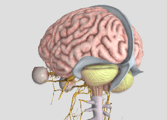
The specific type of brain injury found is a form of herniation called Chiari malformation, where the bottom part of the brain (the cerebellum) drops through the opening in the base of the skull called the foramen magnum. Their findings showed an alarming 23% of the whiplash cases studied had this anatomical abnormality.

Dr. Michael Freeman, Dr. Ezriel Kormel, and colleagues collaborated in this effort and evaluated the patients using MRI in both recumbent (laying down) AND upright positions. Interestingly, they found 5.7% and 5.3% of those in the non-whiplash neck pain group and 9.8% and 23.3% in the whiplash group had the Chiari malformation using the recumbent vs. upright MRI positions, respectfully.

Dr. Kormel stated, “This condition can be quite painful and endanger the patient’s health, with symptoms that may include headaches, neck pain, upper extremity numbness and tingling, and weakness. In a few cases, there can also be lower extremity weakness and brain dysfunction.” In a radio interview, he added the advice that ANYONE suffering from whiplash should see a healthcare provider immediately.

This study is important for a number of reasons. First, it revealed that there is often a more serious injury when whiplash occurs than what is initially found. Second, psychological findings like depression, anxiety, and difficulty coping with the decreased ability or inability to be productive at home or work may suggest the presence of an anatomical injury which simply has not yet been found. Third, MRI is frequently ONLY performed in a laying down position. This study didn’t find much difference between laying vs. weight-bearing MRI positions in the non-whiplash neck pain patients but not so in the whiplash neck pain group! In this group, the ability for MRI to detect Chiari malformation/brain injury more than doubled using weight-bearing MRI.

Expanding the last point, since one out of five whiplash patients had a brain injury that is more likely to be detected using a non-traditional upright MRI position, a “new” standard” for the use of MRI in the evaluation of the whiplash patient should be considered. This is especially important in those cases that are non-responsive to quality care or if their doctor had only ordered a recumbent MRI previously.

Doctors of all disciplines should be aware of this study and the need for a more thorough evaluation, especially when a whiplash patient is not responding as one might expect.

Food Myth Series

01-signs-healthy-fats-mufa.jpg

Over the next few weeks we will be addressing some common food myths that, frankly, drive us a little bit crazy when we hear them. Hopefully we are able to clarify some common misconceptions regarding diet and how what you eat affects how you feel and perform.

We are always open to discussion and want to hear any food myths you have heard in your travels that made you go “What?”.

So, without further delay, food myth #1:

Eating Fats Is Unhealthy

Generations of now adults have been brainwashed into believing that fats = getting fat by the Canada Food Guide. This is simply not the case. While some fats are unhealthy, plenty more have benefits that you won’t find elsewhere. A lower calorie eating plan that includes healthy fats can help people lose more weight than a similar diet that’s low in fat, according to a study in the International Journal of Obesity. Fats make food taste better, keep you fuller and help prevent overeating.

It is true that fats have more calories per gram than proteins and carbs but those calories come with health benefits that the others don’t. Healthy fats like the ones found in salmon, olive oil, nuts and avocados are key to several healthy living goals including:

1. They are a major fuel source for your body (meaning they provides a lot of calories) and are also the main way you store energy.
2. You need fat to help you absorb certain nutrients, such as fat-soluble vitamins (vitamins A, D, E and K) and antioxidants (like lycopene and beta-carotene).
3. Fats are important in giving your cells structure.
4. Omega-3 fats, a type of unsaturated fat, are important for optimum nerve, brain and heart function.

One type of fat you don’t need? Trans fats, an artificial kind of fat found in partially hydrogenated oils and a main ingredient in the food frying process (delicious, yes, but very unhealthy).

Source

So there you go, a quick overview of why fats are important in every healthy diet. Come back next week for more food myth-busting.

health-fats-for-your-heart.jpg


Fibromyalgia and Exercise

295

Are there differences in lifestyle between people with vs. without fibromyalgia (FM)?

A recent study found women with FM found spend more time engaged in sedentary behaviors and less time in physical activity. In the study, researchers followed 413 female patients with FM and 188 age-matched healthy female controls. Researchers used three different approaches to access physical activity: a triaxial accelerometer to examine sedentary time, time spent in physical activity, and step counts.

They discovered those who suffered from FM spent an average of 39 more minutes per day in sedentary activity and 21 fewer minutes per day in light physical activity, 17 fewer minutes per day in moderate physical activity, and 19 fewer minutes per day in moderate-to-vigorous physical activity. In addition, those with FM took a mean of 1,881 fewer steps that those without FM.

Now, this isn’t really a surprise given the fact that people with FM are in pain and more likely to have difficulties sleeping and tolerating prolonged activities. After comparing the sufferers to the non-sufferers, the researchers found only 21% of FM patients vs. 46% of non-FM controls achieved the recommended 150 minutes/week (a little over 20 min. / day) of “moderate-to-vigorous” physical activity. They also found that only 16% vs. 45%, respectively, walked the recommended ≥10,000 steps per day.

One of the BEST forms of exercise for most people is walking. A walking program should be a staple exercise. It’s important to note that this should be GRADUALLY introduced so as to avoid an overuse injury—strain or sprain of the muscles and joints. This gradual introduction into activity is ESPECIALLY important for the FM sufferer as overuse injuries can make them afraid to do something that can REALLY help when done correctly!

Carpal Tunnel Basics

Here it is: carpal tunnel syndrome (CTS) in a nutshell!

WHAT: CTS is caused by an injury to the median nerve (MN) as it travels through the wrist.

WHERE: The eight small carpal bones and a ligament form a tunnel in which tendons and nerves pass through to reach the hand.

HOW: The MN gets pinched/irritated from repetitive stress.

WHY: The tunnel is tight as it includes the MN and nine rapidly moving muscle tendons!

PROGRESS: CTS usually starts slow and often progresses over weeks, months, even years.

SYMPTOMS: Pain, numbness, tingling, and/or weakness of the hand, sparing the little finger.

PROGNOSIS: CTS is easier to treat shortly after it starts, and waiting too long to seek care may lead to worse outcomes.

RISK FACTORS: 1) family history (genetics); 2) women are more likely to suffer from CTS than men; 3) age over 50; 4) manual jobs; 5) pregnancy; 6) conditions like diabetes, hypothyroid, rheumatoid arthritis (RA), osteoarthritis, autoimmune diseases (includes RA, certain types of thyroid disease), gout, kidney disease (especially dialysis patients), Down syndrome, amyloidosis, acromegaly, tumors on the median nerve; 7) medications (those that affect the immune system such as interleukin-2, possibly corticosteroids), anti-clotting drugs such as warfarin, hormone replacement, BCPs; 8) obesity; 9) smoking; 10) alcohol abuse; and 11) trauma/injuries (fractures, tendonitis).

TREATMENT: Ideally, treatment should begin as soon as possible after symptoms first start, but this RARELY occurs due its slow and gradual onset. Non-surgical care includes anti-inflammatory care (ice, anti-inflammatory nutrients—ginger, turmeric, bioflavonoids; NSAIDs like ibuprofen), wrist splinting (primarily at night), corticosteroid injections, job/ergonomic modifications, exercises (yoga, stretching, strengthening, and aerobic fitness), low level laser therapy, ice, acupuncture, and chiropractic care. Chiropractic care includes MANY of the above PLUS manual therapies applied to the neck, shoulder, arm, wrist, and hand.

Nutshell

Trigger points in the sternocleidomastoid muscle. By Bryan Cobb RMT.

Screen Shot 2017-12-01 at 10.39.50 AM.png

The sternocleidomastoid muscle or “scm” is a strap like muscle located in the front of the neck. It originates on the manubrium of the sternum and the medial clavicle, and inserts on the mastoid process of the temporal bone( the lump or bone behind the ear). When working together the scm flexes the head and neck. The actions of one muscle working by itself is to rotate the face to the opposite side and lift it toward the ceiling.

Trigger points in this muscle usually form due to acute injury or to chronic postural stress. These trigger points can cause a wide variety of symptoms including pain felt in the cheek, temple, forehead, top of the head, and base of the skull. Trigger points in these muscles can also refer deep into the ear and cause postural dizziness.

Hip or Low Back?

Lumbar and Pelvis

Technically, the hip is the ball-and-socket joint between the long bone of the thigh and the pelvis; but more often than not, many people will point to a number of different places on their back or pelvis and say, “My hip is killing me” when it’s not really “the hip” at all!
Hip pain can be located in the front (groin area), the side, the back, or in the buttocks. The “classic sign” of hip pain is reproduced most consistently when you try to cross your legs—put your ankle on the far end of the thigh and then push down on your knee. This may feel tight and cause pain in the groin area. For many people, hip pain is also reproduced when they cross their legs and then pull their knee towards the opposite shoulder.
The hip is a VERY strong joint due to the deep receiving cup of the pelvis and the round ball that fits nicely into it. Because it’s a freely moving joint, there is a joint capsule. The capsule is lined with tissue that produces an oily substance that lubricates the joint (called synovial fluid), and when hip pain occurs, this can be caused by a capsulitis (inflammation of the capsule) with a buildup of synovial fluid (called synovitis).
When the smooth, shiny surface of the ball starts to wear thin (which can eventually wear away down to the bone), that’s a condition called “osteoarthritis.” This occurs over time for many and may eventually result in the need for a hip replacement. This usually isn’t needed until an individual is in their late 60s or older (if at all), but for those who injure a hip earlier in life, the “wearing out” process may accelerate and a hip replacement may be needed well before old age.
There are many studies that report low back and hip arthritis often occur together, and differentiating between the two can sometimes be a challenge. For example, pain can radiate from the hip to the knee, which many doctors will diagnose as “sciatic nerve.” But hip pain can present exactly the same, making it hard to determine if it’s low back-generated pain or hip-generated pain.
This is why it’s SO IMPORTANT that your doctor of chiropractic conduct a careful history and examination. There are specific tests that he or she will perform that help determine which of the two is causing the pain. There are times when they may find BOTH problems co-existing together, making it necessary to manage two problems, rather than just one.
There are many mobilization, manipulation, soft tissue therapies, modalities, and exercises available to patients with both hip and low back pain. So if you’re not sure what is bothering you and you don’t know what to do, visit your doctor of chiropractic and he or she will help you through this.

The Best Diet For Fibromyalgia?

Anti-Inflam

Fibromyalgia (FM) and its cause remains a mystery, but most studies suggest that FM is NOT the result of a single event but rather a combination of many physical, chemical, and emotional stressors.

The question of the month regarding the BEST FM diet is intriguing since one might assume that the many causes should mean that there isn’t one dietary solution. But is that true? Could there be a “best diet” to help ease the symptoms from such a multi-faceted disorder?

Certainly, healthy eating is VERY important for ALL of us regardless of our current ailment(s). Obesity is rampant largely due to the fact that 60% of the calories consumed by the “typical” American center around eating highly inflaming food that include those rich in Sugar, Omega-6 oil, Flour, and Trans fats (“SOFT” foods, if you will!). Obesity has been cited as “an epidemic” largely due to kids and adults becoming too sedentary (watching TV, playing on electronic devices, etc.) and eating poorly.

Perhaps the BEST way to manage the pain associated with FM and to maintain a healthy BMI (Body Mass Index, or ratio between height and weight) is to substitute ANTI-INFLAMING foods for those that inflame (or SOFT foods).

You can simplify your diet by substituting OUT “fast foods” for fruits, vegetables, and lean meats. So there you have it. It’s that simple. The problem is making up your mind to change and then actually doing it. Once these two things take place, most everyone can easily “recalibrate” their caloric intake and easily adapt.

Not only have studies shown that chronic illnesses like heart disease, stroke, and diabetes significantly benefit by following this simple dietary shift, but so does pain arising from the musculoskeletal system! This is because the human body is made up largely of chemicals, and chemical shifts are constantly taking place when it moves. If you reach for an anti-inflammatory drug like ibuprofen or naproxen and it helps, it’s because you ARE inflamed and the drug reduces the pain associated with that inflammation. This is an indication that an anti-inflammatory diet WILL HELP as well (but without the negative side effects)!

The list of chronic conditions that result in muscle pain not only includes FM but also obesity, metabolic syndrome, and type II diabetes. Conditions like tension-type and migraine headaches, neck and back pain, disk herniation, and tendonopathies and MANY more ALL respond WELL to making this SIMPLE change in the diet. For more information on how to “DEFLAME,” visit http://www.deflame.com! It could be a potential “lifesaver!”

I get dizzy when I have a headache. Should I worry?

Vertigo_view_03,_Andels_Hotel_Łódź.jpg

Dizziness, neck pain, and headaches are very common symptoms that may or may not occur at the same time. Though this interrelationship exists, this month’s article will focus primarily on dizziness, particularly related to dizziness that occurs after standing.

First, it is important to point out that it is VERY common to be light headed or dizzy when standing up too fast, which is typically referred to as orthostatic hypotension (OH). OH is frequently referred to as a benign symptom, but new information may challenge this thought.

Let’s review what happens. When we are lying down, our heart does not have to work as hard as when we are upright; therefore, our blood pressure (BP) is usually lower while we lay in bed. When standing up, blood initially pools in the legs until an increase in blood pressure brings oxygen to the brain. This either resolves or prevents dizziness.

Orthostatic hypotension is defined as a blood pressure drop of >20 mm Hg systolic (the upper number—heart at FULL contraction), 10 mm Hg diastolic (lower number—heart at FULL rest), or both. This typically occurs within seconds to a few minutes after rising to a standing position.

There are two types of OH—delayed OH (DOH) where the onset of symptoms are not immediate but occur within three minutes of standing and “full” OH, which is more serious and occurs immediately upon rising. According to a 2016 study published in the prestigious journal Neurology, researchers reviewed the medical records of 165 people who had undergone autonomic nervous system testing for dizziness. The subjects averaged 59 years of age, and 48 were diagnosed with DOH, 42 with full OH, and 75 subjects didn’t have either condition.

During a ten-year follow-up, 54% of the DOH group progressed to OH, of which 31% developed a degenerative brain condition such as Parkinson’s disease or dementia. Those with initial DOH who also had diabetes were more likely to develop full OH vs. those without diabetes.

The early death rate in this 165 patient group was 29% for those with DOH, 64% with full OH, vs. 9% for those with neither diagnosed condition. The authors point out that those initially diagnosed with DOH who did NOT progress into full OH were given treatment that may have improved their blood pressure.

The authors state that a premature death might be avoided by having DOH and OH diagnosed and properly managed as early as possible. They point out that a prospective study is needed since this study only looked back at medical records of subjects who had nervous system testing performed at a specialized center, and therefore, these findings may not apply to the general population.

The value of this study is that this is the FIRST time a study described OH (or DOH) as a potentially serious condition with recommendations NOT to take OH/DOH lightly or view it as a benign condition. Since doctors see this a lot, a closer evaluation of the patient is in order.

Do You Have CTS?

Carpal tunnel syndrome (CTS) can be an extremely painful and activity-limiting condition. It affects many people of all ages and genders, though women are affected more often than men. But how do you know if what you are suffering from is truly CTS or if it’s another condition that’s producing the symptoms in your hand or wrist?

Carpal tunnel syndrome occurs when the median nerve is compressed as it passes through the wrist. However, the median nerve travels out of the neck, through the shoulder, elbow, and forearm before it passes through the wrist and into the hand. Pinching of the median nerve ANYWHERE along its course can give rise to the signs and symptoms of CTS including numbness, tingling, and/or pain into the hand and index, third, and thumb-side half of the fourth digits, and sometimes the thumb. If the pinch is significant enough, weakness can also occur. Sometimes the median nerve can become compressed at both the wrist and other body sites as it travels from the spinal cord to your hand, that’s why it’s important for a doctor to check for impingements along the entire course of the nerve.

But compression of the median nerve isn’t the only thing that can produce symptoms in the hand. Here are a couple of the more common conditions that are often confused with CTS:

1)  Ulnar neuropathy: This is pinching of the ulnar nerve (at the neck, shoulder, elbow, or wrist) but this gives rise to a similar numbness/tingling BUT into the pinky-side of the fourth and the fifth fingers (not the thumb-side of the hand). The most common pinch location is either at the neck or the inner elbow, the latter of which is called “cubital tunnel syndrome” or CuTS.

2)  Tendonitis: There are a total of nine tendons that pass through the carpal tunnel that help us grip or make a fist. Similarly, there are five main tendons on the back side of the hand that allow us to open our hands and spread our fingers. ANY of these tendons can get strained or torn, which results in swelling and pain as well as limited function BUT there is usually NO NUMBNESS/TINGLING!

3)  DeQuervain’s disease: This is really a tendonitis of an extensor tendon of the thumb and its synovial sheath that lubricates it resulting in a “tenosynovitis.” This creates pain with thumb movements, especially if you grasp your thumb in the palm of your hand and then bend your wrist sideways towards the pinky-side of the hand.

Chiropractors are well-trained to diagnose and treat patients with CTS. And if you don’t have CTS but another condition listed above, they can offer treatment (or a referral, if necessary) to help resolve it so you can return to your normal activities as soon as possible.

DOn't know

What is Facet Syndrome?

As stated in last week’s article, low back pain (LBP) can arise from many different structures. Lumbar facet syndrome is one that involves the facet joint and includes both acute (new) and chronic (old) varieties. The facet joint is synonymous with the zygapophyseal joint, so if you hear that word, don’t let it throw you off! Approximately 45% of patients with chronic low back pain suffer from “facet syndrome” (FS) in which the facets are the low back pain generator.
There are many conditions that give rise to FS. Some of these include the straining of the surrounding joint capsule (the capsule holds the joint securely together), joint hypomobility (reduced motion in the joint), a synovial cyst (similar to a ganglion on the back of the wrist but its located inside the joint), and degeneration (also called osteoarthritis—the wearing out type of arthritis).
Because facet syndrome can accompany other conditions, a doctor of chiropractic must evaluate each patient individually and manage each person appropriately. In “pure” facet syndrome, pain rarely ever passes the level of the knee and does not cause neurological loss (weakness, loss of reflex, etc.). It can create numbness but usually NOT beyond the knee. Pain is usually not worsened by hip movements such as straight leg raise or hip rotation.
The facet joint’s “job” (at least in part) is to limit or guard twisting movements in the upper lumbar/low back region, and the lower lumbar facets are shaped to limit motion when bending forwards and backwards. Facet joints are unique because they are innervated by specific nerves that can be blocked by injecting an anesthetic agent to determine if the facet (and its innervating nerve) is the main source of pain. The surrounding capsule around the facet joint contains mechanoreceptors (cells that detect movement) and nociceptors (cells that detect pain) that fire when the facet joint is compressed/jammed or over-stretched. These nociceptors can become “hypersensitized” (very irritable) when they remain inflamed over time.
In many patients, injury to a facet joint is the result of many microtraumas over a period of time and not one single isolated event. For example, repeatedly bending backwards, twisting, and leaning to one side can stretch the joint capsule and fatigue it until some capsular tissues finally “give” and it inflames which generates pain.
These joints commonly become arthritic with age, which is one reason people over 50-60 years old commonly present with FS. Osteoarthritis results in a narrowing of the joint space and causes a more permanently “jammed” joint. This is one reason many elderly people walk partially bent over—as bending forwards opens the facet joints and “feels good!”
facet joint
The good news is that chiropractic manipulation is a highly effective treatment for facet syndrome, and most patients feel much better within the first or second week of care (often within three to five visits).