PFPS Cont. You want details?

Screen Shot 2017-10-31 at 11.15.32 AM.png

The muscles of the hip provide not only local stability, but also play an important role in spinal and lower extremity functional alignment. (1-4) While weakness in some hip muscles (hip extensors and knee extensors) is well tolerated, weakness or imbalance in others can have a profound effect on gait and biomechanical function throughout the lower half of the body. (5) Weakness of the hip abductors, particularly those that assist with external rotation, has the most significant impact on hip and lower extremity stability. (5,6)

Hip Abduction.gif

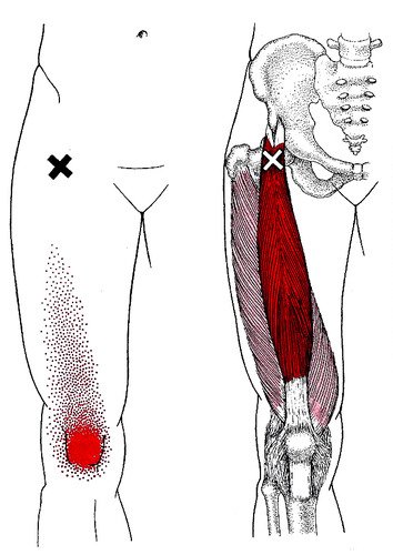
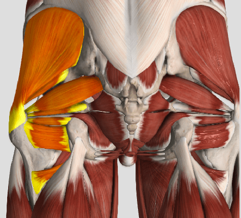
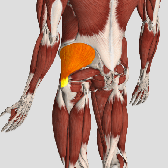
The gluteus medius is the principal hip abductor. When the hip is flexed, the muscle also assists the six deep hip external rotators (piriformis, gemelli, obturators, and quadratus femoris). The gluteus medius originates on the ilium just inferior to the iliac crest and inserts on the lateral and superior aspects of the greater trochanter. While the principal declared action of the gluteus medius is hip abduction, clinicians will appreciate its more valuable contribution as a dynamic stabilizer of the hip and pelvis- particularly during single leg stance activities like walking, running, and squatting. The gluteus medius contributes approximately 70% of the abduction force required to maintain pelvic leveling during single leg stance. The remainder comes predominantly from 2 muscles that insert onto the iliotibial band: the tensor fascia lata and upper gluteus maximus.  Hip abductor strength is the single greatest contributor to lower extremity frontal plain alignment during activity. (6)

Screen Shot 2017-10-31 at 11.12.00 AM

Incompetent hip abductors and/or external rotators allows for excessive adduction and internal rotation of the thigh during single leg stance activities. This leads to a cascade of biomechanical problems, including pelvic drop, excessive hip adduction, excessive femoral internal rotation, valgus knee stress, and internal tibial rotation. (1,7-12)


References
1. Szu-Ping Lee, Powers C. Description of a Weight-Bearing Method to Assess Hip Abductor and External Rotator Muscle Performance. JOSPT. Volume 43, Issue 6
2. Crossley KM, Zhang WJ, Schache AG, Bryant A, Cowan SM. Performance on the single-leg squat task indicates hip abductor muscle function. Am J Sports Med. 2011;39:866-873.
3. Presswood L, Cronin J, Keogh JWL, Whatman C. Gluteus medius: applied anatomy, dysfunction, assessment, and progressive strengthening. Strength Cond J. 2008;30:41-53.
4. Sled EA, Khoja L, Deluzio KJ, Olney SJ, Culham EG. Effect of a home program of hip abductor ex- ercises on knee joint loading, strength, function, and pain in people with knee osteoarthritis: a clinical trial. Phys Ther. 2010;90:895-904.
5. van der Krogt MM, Delp SL, Schwartz MH How robust is human gait to muscle weakness? Gait Posture. 2012 Feb 29.
6. Laurie Stickler, Margaret Finley, Heather Gulgin Relationship between hip and core strength and frontal plane alignment during a single leg squat Physical Therapy in Sport Available online 2 June 2014
7. Ireland ML, Willson JD, Ballantyne BT, Davis
IM. Hip strength in females with and without patellofemoral pain. J Orthop Sports Phys Ther. 2003;33:671-676.
8. Noehren B, Davis I, Hamill J. ASB clinical biome- chanics award winner 2006: prospective study of the biomechanical factors associated with iliotib- ial band syndrome. Clin Biomech (Bristol, Avon). 2007;22:951-956.
9. Powers CM. The influence of abnormal hip me- chanics on knee injury: a biomechanical perspec- tive. J Orthop Sports Phys Ther. 2010;40:42-51.
10. Powers CM. The influence of altered lower- extremity kinematics on patellofemoral joint dysfunction: a theoretical perspective. J Orthop Sports Phys Ther. 2003;33:639-646.
11. Sigward SM, Powers CM. Loading characteristics of females exhibiting excessive valgus moments during cutting. Clin Biomech (Bristol, Avon). 2007;22:827-833
12. Souza RB, Powers CM. Differences in hip kine- matics, muscle strength, and muscle activation between subjects with and without patellofemo- ral pain. J Orthop Sports Phys Ther. 2009;39:12- 19.

Chiro & Concussions

head-shot2

Whiplash Associated Disorders (WAD) is the appropriate terminology to use when addressing the myriad of symptoms that can occur as a result of a motor vehicle collision (MVC). In a recent publication in The Physician and Sports Medicine (Volume 43, Issue 3, 2015; 7/3/15 online:1-11), the article “The role of the cervical spine in post-concussive syndrome” takes a look at the neck when it’s injured in a car accident and how this relates to concussion.

It’s estimated about 3.8 million concussion injuries, also referred to as “mild traumatic brain injury” (mTBI), occur each year in the United States. Ironically, it’s one of the least understood injuries in the sports medicine and neuroscience communities. The GOOD NEWS is that concussion symptoms resolve within 7-10 days in the majority of cases; unfortunately, this isn’t the case with 10-15% of patients. Symptoms can last weeks, months, or even years in this group for which the term “post-concussive syndrome” (PCS) is used (defined as three or more symptoms lasting for four weeks as defined by the ICD-10) or three months following a minor head injury (as defined by the Diagnostic and Statistical Manual of Mental Disorders).

There have been significant advances in understanding what takes place in the acute phase of mTBI, but unfortunately, there is no clear physiological explanation for the chronic phase. Studies show the range of force to the head needed to cause concussion is between 60-160g (“g” = gravity) with 96.1g representing the highest predictive value in a football injury, whereas as little as 4.5g of neck acceleration can cause mild strain injury to the neck. In spite of this difference, the signs and symptoms reported by those injured in low-speed MVCs vs. football collisions are strikingly similar!

Research shows if an individual sustains an injury where the head is accelerated between 60-160g, it is HIGHLY likely that the tissues of the cervical spine (neck) have also reached their injury threshold of 4.5g. In a study that looked at hockey players, those who sustained a concussion also had WAD / neck injuries indicating that these injuries occur concurrently. Injuries to the neck in WAD include the same symptoms that occur in concussion including headache, dizziness/balance loss, nausea, visual and auditory problems, and cognitive dysfunction, just to name a few.

The paper concludes with five cases of PCS that responded well to a combination of active exercise/rehabilitation AND passive manual therapy (cervical spine manipulation). The favourable outcome supports the concept that the neck injury portion of WAD is a very important aspect to consider when treating patients with PCS!

This “link” between neck injury and concussion explains why chiropractic care is essential in the treatment of the concussion patient! This is especially true when the symptoms of concussion persist longer than one month!

We realize you have a choice in whom you consider for your health care provision and we sincerely appreciate your trust in choosing our service for those needs.  If you, a friend, or family member requires care for Whiplash, we would be honoured to render our services.

Preventing Injury In Young Athletes

Identifying and reducing risk factors in sports in especially important in young athletes. A review published in “The Adolescent Athlete” journal found that up to 50% go injuries could be avoided with preventative measures for youth participating in sport. Some key items to consider when looking at preventing injuries include:

  • A pre-season screening program to identify muscle imbalances, weakness, previous injury locations and progress of healing at those sites. Waiting for an injury in-season is cuter productive for all athletes but especially young ones.
  • An off-season general strength and conditioning program. This should be aimed at maximizing an athletes general movement skills, sport specific injury prevention through balance and strength and general mobility.
  • Awareness of how growth affects athletes from not only a performance point of view but also how growth impacts muscles, strength and co-ordination, especially in the lower limbs.
  • Awareness of how specific skill sets impact the body through repetitive stress and how to prevent that stress through load management, practice balance and body awareness.
  • Early intervention is always the best course of action when dealing with a young athlete. At first sign of dysfunction, even without pain a professional should be consulted to ensure prevention of exacerbation on the condition. “Toughing it out” leads to more issues down the line.
  • Understanding that pain is not a “normal part of sport”. The old adage “No pain, no gain” needs to be scrubbed from the sporting world. Play and practice smarter and pain can be avoided in all sports.

If you are a coach, parent or athlete looking for more information on these or any sports injury related items, feel free to contact us at any time. 

Why trigger point therapy?

People often think of a massage as a relaxing experience, something they might do occasionally, or give as a gift along with a trip to the spa. Trigger point massage therapy is another kind of massage used to treat pain and physical dysfunction. Trigger points can develop in people from all walks of life. They can affect people of all ages, office workers and labourers, elite and weekend athletes, post surgical patients, people with acute pain from injury and people with chronic pain. Trigger point massage therapy can treat a wide variety of physical conditions such as:

– Migraines

– back pain.

– sciatica

– Carple tunnel syndrome

– achy persistent pain

– pain from Fibromyalgia

– post surgical pain and scarring

– soft tissue injuries related to sports

– TMJ dysfunction

Myofascial trigger points and pain.

TP’s were first brought to the attention of the medical world by Dr. Janet G. Travell. Dr. Travell, physician to President John F. Kennedy, is the acknowledged Mother of Myofascial Trigger Points. In fact, “Trigger Point massage, the most effective modality used by massage therapists for the relief of pain, is based almost entirely on Dr. Travell’s insights.”2 Dr. Travell’s partner in her research was Dr. David G. Simons, a research scientist and aerospace physician.

Trigger Points are very common. In fact, Travell and Simons state that TP’s are responsible for, or associated with, 75% of pain complaints or conditions.1 With this kind of prevalence, it’s no wonder that TP’s are often referred to as the “scourge of mankind”.

Trigger Points can produce a wide variety of pain complaints. Some of the most common are migraine headaches, back pain, and pain and tingling into the extremities. They are usually responsible for most cases of achy deep pain that is hard to localize.

A TP will refer pain in a predictable pattern, based on its location in a given muscle. Also, since these spots are bundles of contracted muscle fibres, they can cause stiffness and a decreased range of motion. Chronic conditions with many TP’s can also cause general fatigue and malaise, as well as muscle weakness.

Gluteus medius trigger points.

The Gluteus medius is located in your hip. It is important for hip stability, and functions to abduct the hip. When trigger points form in this muscle pain can be felt deep into the sacrum and the hip, as well as down the lateral thigh. Trigger points in this muscle are an often overlooked source of low back and sacral pain.

Planter fascitis and trigger points

Planter fascitis is a painful condition affecting the bottoms of the feet. It involves inflammation and tightness of the planter fascia, which is a tough layer of connective tissue on the bottoms of the feet. Trigger points in the calf and foot muscles are a leading cause of planter fascitis. These points not only cause the muscle to become tight which in turn causes the fascia to be tight, but the trigger point pain referral patterns of these muscle will cause pain to be felt in the bottoms of the feet and heal. These causes are often overlooked leading to ineffective treatment and prolonged suffering.

Trigger points in the gluteus medius.

The gluteus medius muscle is located in the hip. It is a major pelvic stabilizer during walking and running. It is also a main abductor of the hip. Trigger points in this muscle will refer pain deep into the sacrum and s.i. joints. Pain will also be refered into the buttock as well as into the low back along the belt line.

Trigger points in the Q.L. Muscle

the quadratus lumborum muscle or “q.l.” Is a muscle located in your lower back. It originates on the iliac crest and iliolumbar ligament, and interns onto the last rib and transverse processes of the lumbar vertebrae. This muscle functions to stabilize the lumbar spine as well as laterally flex the spine. It will also hike the hip. Acting bilaterally it will extend the lumbar spine. Trigger points will often develop in this muscle. Trigger point referral will produce pain in the S.I. Joint and the lateral hip as well as the buttock. The pain referred into the S.I. Joint is often misdiagnosed as S.I. Joint dysfunction.

The rectus femoris muscle is one of your quad muscles. It acts primarily to extend the knee but it also helps to flex the hip. This muscle is often overloaded from athletic activity, but it also can become chronically shortened from prolonged sitting. Trigger points will refer pain deep into the knee producing a deep ache felt into the joint.