Choosing to walk the golf course is an excellent way to increase your cardiovascular activity during the summer. However, walking 18 holes equates to roughly four to five miles of continuous walking, often on uneven terrain, in variable weather conditions, and while carrying or pushing equipment. This physical load can quickly fatigue the structures of the feet and ankles.
Common Foot and Ankle Stressors
The repetitive stress of walking the course can lead to several acute, localized issues if the lower body is not properly supported or given adequate time to recover:
Plantar Fasciitis Flare-Ups: The thick band of tissue running across the bottom of your foot can become inflamed, causing sharp heel pain, especially during the first few steps of the morning.
Achilles Tendon Strain: Walking up steep tee boxes or out of deep bunkers places heavy eccentric loads on the Achilles, leading to micro-tearing and stiffness.
Ankle Sprains: Uneven lies, hidden sprinkler heads, and thick rough present constant tripping hazards that can easily roll an unprepared ankle.
Post-Round Recovery Tactics
To maintain your lower body health throughout the golf season, prioritize active recovery as soon as your round ends:
Active Stretching: Focus on stretching the calf muscles (both the gastrocnemius and soleus) to relieve tension on the Achilles tendon and the plantar fascia.
Ice Application: If you notice localized heat or swelling around the ankle joint after a round, apply ice for 15-20 minutes to manage the inflammatory response.
Footwear Assessment: Ensure your golf shoes provide adequate arch support and torsional stability. Shoes that are overly flexible can contribute to foot fatigue over 18 holes.
Keep your feet course-ready. If foot or ankle pain is altering your gait or keeping you off the course, our clinical team can help.
Booking: Same-day or next-day appointments are available.
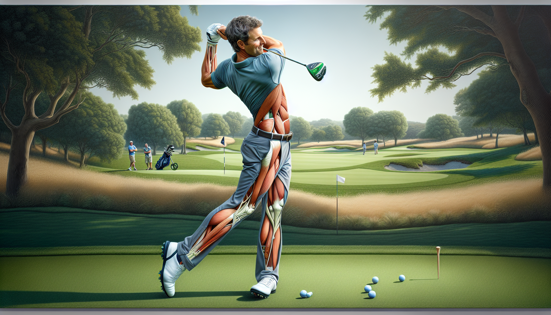
For the dedicated golfer, maximizing performance means more than just hitting the range. The physical load of playing two to three rounds a week adds significant strain to the back, hips, and joints. Implementing structured recovery strategies is just as important as perfecting your swing mechanics.
Managing the Physical Load
Balancing frequent rounds of golf alongside a standard gym routine—such as a three-day lifting split—requires careful load management. The repetitive rotational force of the golf swing takes a toll, especially when utilizing heavier, stiff-flex steel shafts, like 120-gram X-flex models, which demand more physical exertion to load properly. Spacing out your heavy gym days from your tee times ensures your muscles have time to repair.
Tracking Performance as a Recovery Metric
Monitoring technical performance data is not just for improving your handicap; it is an excellent tool for tracking recovery. If you utilize a launch monitor for indoor practice, keep an eye on your baseline metrics. A sudden, unexplained drop in clubhead speed or ball speed often points to physical fatigue or inadequate muscle recovery rather than a sudden mechanical flaw.
Proactive Tissue Management
Walking 18 holes multiple times a week demands a lot from your lower body. Proactive recovery should include:
Hydration: Maintaining fluid levels to keep tissues pliable and reduce cramping.
Mobility Work: Focusing on hip internal and external rotation, as well as thoracic spine mobility, on your non-playing days.
Active Recovery: Engaging in light, low-impact movement (like walking or cycling) on off-days to promote blood flow without adding stress to the joints.
Ignoring early signs of stiffness can lead to compensatory swing habits, which often result in overuse injuries in the lower back or elbows.
Keep your body course-ready. If a nagging strain is impacting your game, River East Minor Injury Clinic can help you address it before it sidelines your season.
Booking: Same-day or next-day appointments are available.
When you sustain a minor injury—whether it is a rolled ankle on a morning run, a strained lower back from lifting, or a wrist injury from a weekend tournament—prompt care is essential. However, the prospect of waiting for hours in a crowded, uncomfortable clinic waiting room often deters people from seeking the immediate professional assessment they need.
At River East Minor Injury Clinic, we have implemented a streamlined, scheduled approach to minor injury care. Here is exactly how our same-day and next-day booking system works.
The Shift from “Walk-In” to “Scheduled” Care
Traditional walk-in models are designed to triage a massive variety of unpredictable health concerns, from minor illnesses to severe infections. Because we exclusively treat minor physical injuries—and do not treat illnesses like the flu or chronic medical conditions—we can accurately predict our clinical flow.
By utilizing a dedicated online scheduling system, we eliminate the uncertainty of the waiting room. You receive a guaranteed appointment time, allowing you to rest comfortably at home until it is time to be seen by our team.
Step-by-Step: Securing Your Appointment
Visit Our Online Portal: Head to our website at http://www.rivereastminorinjury.ca. Our booking platform is accessible 24/7 from your smartphone or computer.
Select Your Time: Choose from our available same-day or next-day appointment slots that fit your schedule.
Provide Preliminary Details: You will be prompted to fill out a brief, secure intake form detailing the nature of your injury. This allows our clinical team to prepare for your specific needs before you even arrive.
Receive Confirmation: Once booked, you will receive a clear confirmation of your time and directions to our facility.
What to Expect When You Arrive
When you arrive at 1191 Rothesay Street for your scheduled appointment, our goal is to move you directly into the assessment phase.
You will be evaluated by our multidisciplinary team, which may include registered nursing assessment, physiotherapy, and chiropractic care. Because your time is reserved exclusively for you, our practitioners can conduct a thorough, unhurried physical examination. We will explain your diagnosis clearly, discuss the evidence-based treatment options available, and work with you to develop a customized recovery plan.
Efficient Care for an Active Community
An unexpected sprain or minor fracture disrupts your daily life. The process of getting it assessed should not cause further frustration. By offering convenient same-day online booking, River East Minor Injury Clinic ensures that Winnipeg residents have access to prompt, professional musculoskeletal care exactly when they need it most.
Whether you are stepping awkwardly off a curb, tweaking your lower back on the golf course, or rolling an ankle during a weekend hockey game, sudden physical setbacks happen. When they do, you are often left wondering: Is this an emergency, or is it something I can just walk off?
At River East Minor Injury Clinic, we aim to bridge the gap between standard home care and the hospital emergency room. To help you make the best decision for your health, let us break down exactly what constitutes a “minor” injury and explore the key differences between two of the most common issues we treat: sprains and strains.
Defining a “Minor” Injury
In the medical field, a minor injury refers to a non-life-threatening physical trauma that involves the musculoskeletal system—meaning your bones, joints, muscles, ligaments, and tendons. These are the acute injuries that cause immediate pain and limit your mobility, but do not require complex emergency interventions like surgery or advanced trauma care.
Examples of minor injuries include:
Sprains and strains
Minor fractures (possibly broken bones that have not pierced the skin)
Sports-related joint injuries
Workplace or Motor Vehicle Accidents
What is NOT a minor injury? It is equally important to understand what a minor injury clinic does not handle. We are exclusively dedicated to physical injuries. We do not treat illnesses. If you are experiencing symptoms like a fever, cough, cold, flu, or an infection, you should seek care from your primary care provider, an urgent care centre, or a minor illness clinic.
Sprains vs. Strains: What is the Difference?
People often use the words “sprain” and “strain” interchangeably, but they actually refer to damage to two entirely different types of soft tissue in the body.
The Sprain (Ligament Damage)
A sprain occurs when you stretch or tear a ligament. Ligaments are the tough, fibrous bands of tissue that connect bone to bone, acting as the stabilizing anchors for your joints.
How it happens: Sprains typically occur from sudden twisting motions, pivoting, or landing awkwardly. The classic example is a rolled ankle, but wrist and knee sprains are also incredibly common.
Symptoms: You will generally experience immediate pain, localized swelling, bruising, and a noticeable restricted range of motion. You might even hear a “pop” at the moment of injury.
The Strain (Muscle or Tendon Damage)
A strain, on the other hand, involves the stretching or tearing of a muscle or a tendon. Tendons are the thick cords of tissue that connect your muscles to your bones.
How it happens: Strains are often the result of sudden, heavy lifting, overstretching, or explosive movements. Pulling a hamstring while sprinting or throwing out your lower back while doing yard work are classic strains.
Symptoms: Strains are characterized by muscle spasms, cramping, weakness in the affected area, swelling, and sharp pain when attempting to move the muscle.
Why You Shouldn’t “Just Walk It Off”
When a sprain or strain happens, the standard advice is often to apply ice and rest. While the R.I.C.E. method (Rest, Ice, Compression, Elevation) is a great first step, trying to tough out a musculoskeletal injury without professional assessment can lead to long-term issues.
Without a proper diagnosis, you might be walking on a minor fracture disguised as a sprain, or you might develop compensatory movement habits that lead to chronic joint instability. Getting a prompt, professional assessment allows you to understand the exact nature of the damage and begin a targeted recovery plan immediately.
Same Day or Next Day Care
River East Minor Injury Clinic was designed to provide you with rapid, professional care on your schedule.
Our Nurse Practitioner is here to assess, diagnose, and treat your sprains and strains efficiently. We offer scheduled same-day and next-day appointments, meaning you get the focused care you need exactly when you need it, with zero walk-in waiting.
Ready to start your recovery? 📍 Find us at: 1191 Rothesay Street, Winnipeg
River East Minor Injury Clinic is officially open!
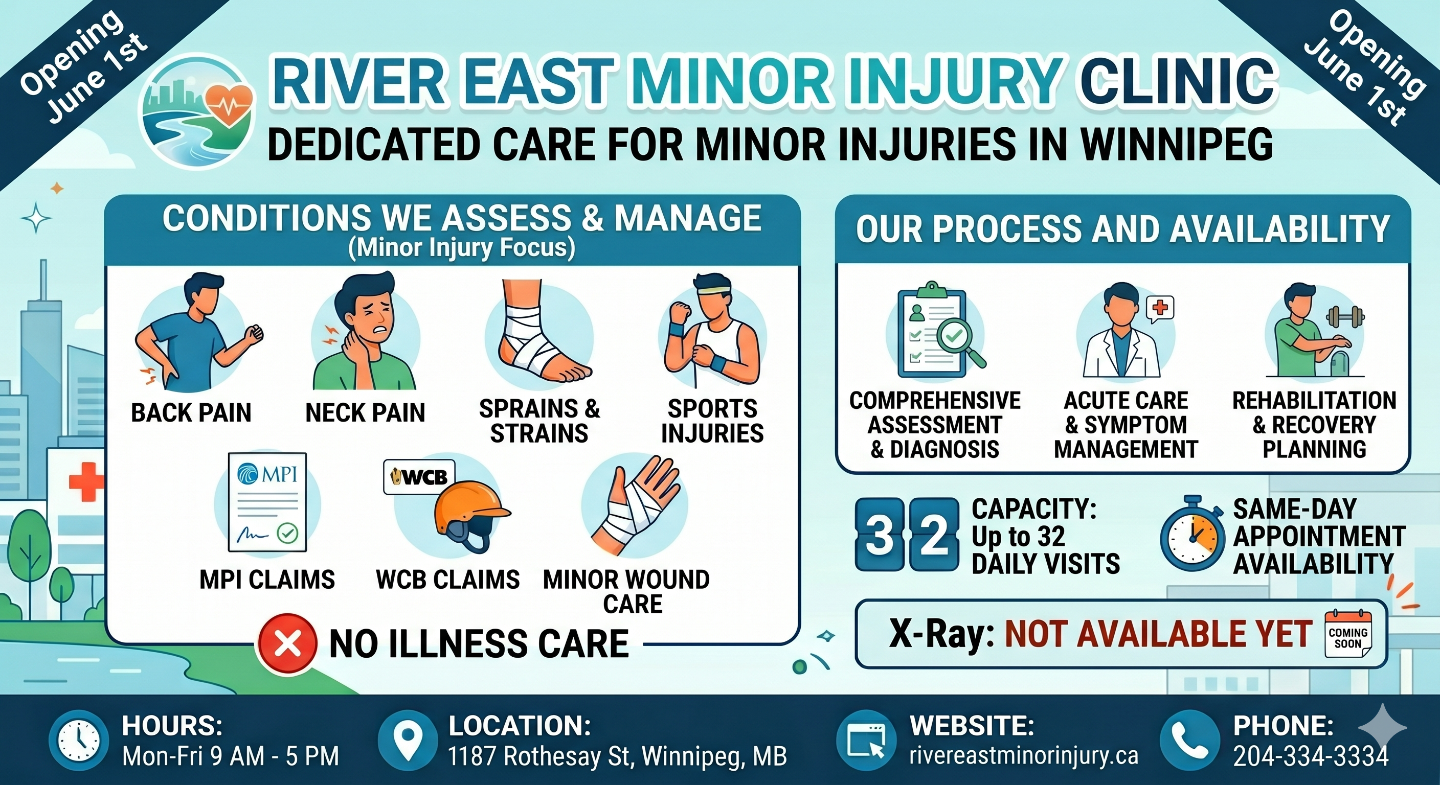
We provide dedicated, multidisciplinary assessment and treatment for minor physical injuries. If you have experienced a recent sprain, strain, minor fracture, or sports-related injury, our team of healthcare professionals is here to help you begin your recovery.
What we treat:
Sprains and strains
Minor fractures
Sports and activity injuries
Workplace and Motor Vehicle Injuries
What we do not treat:(Please visit your primary care provider, an emergency room, or a minor illness clinic for the following)
Coughs, colds, or flu
Fevers or infections
Chronic health conditions or illnesses
To ensure you receive timely care without the uncertainty of walk-in wait times, we offer scheduled same-day and next-day appointments.
Your low back consists of 5 individual vertebrae stacked on top of each other. Flexible cushions called “discs” live between each set of vertebrae. A disc is made up of two basic components. The inner disc, called the “nucleus”, is like a ball of jelly about the size of a marble. This jelly is held in place by the outer part of the disc called the “annulus”, which is a tough ligament that wraps around the inner nucleus much like a ribbon wrapping around your finger. Your low back relies on discs and other ligaments for support. “Discogenic Low Back Pain” develops when these tissues are placed under excessive stress, much like a rope that frays when it is stretched beyond its normal capacity. Most commonly, disc pain is not the result of any single event, but rather from repeated overloading. Your lumbar discs generally manage small isolated stressors quite well, but repetitive challenges lead to injury in much the same way that constantly bending a piece of copper wire will cause it to break. Examples of these stressors include: bad postures, sedentary lifestyles, poor fitting workstations, repetitive movements, improper lifting, or being overweight.
Approximately one third of adults will experience pain from a lumbar disc at some point in their lifetime. The condition is more common in men. Most lumbar disc problems occur at one of the two lowest discs- L5 or L4. Smokers and people who are generally inactive have a higher risk of lumbar disc problems. Certain occupations may place you at a greater risk, especially if you spend extended periods of time sitting or driving. People who are tall or overweight have increased risk of disc problems.
Symptoms from disc pain may begin abruptly but more commonly develop gradually. Symptoms may range from dull discomfort to surprisingly debilitating pain that becomes sharper when you move. Rest may relieve your symptoms but often leads to stiffness. The pain is generally centered in your lower back but can spread towards your hips or thighs. Be sure to tell your doctor if your pain extends beyond your knee, or if you have weakness in your lower extremities or a fever.
Repeated injuries cause your normal healthy elastic tissue to be replaced with less elastic “scar tissue.” Over time, discs may dehydrate and thin. This process can lead to ongoing pain and even arthritis. Patients who elect to forego treatment and “just deal with it” develop chronic low back pain more than 60% of the time. Seeking early and appropriate treatment like the type provided in our office is critical.
Depending on the severity of your injury, you may need to limit your activity for a while, especially bending, twisting, and lifting, or movements that cause pain. Bed rest is not in your best interest. You should remain active and return to normal activities as your symptoms allow. Light aerobic exercise (i.e. walking, swimming, etc) has been shown to help back pain sufferers. The short-term use of a lumbar support belt may be helpful. Sitting makes your back temporarily more vulnerable to sprains and strains from sudden or unexpected movements. Be sure to take “micro breaks” from workstations for 10 seconds every 20 minutes.
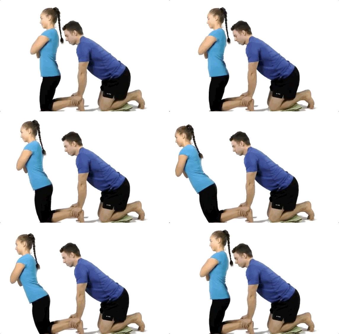
A great injury prevention movement is the glute-ham raise. Done after a warm up and prior to competition it will significantly reduce the odds of hamstring strains in running athletes in sports like Soccer, Football and Sprinting.
To perform the movement:
Begin in a tall kneeling position on a cushion or pillow.
Partner grabs and holds ankles to ground or hook your feet under a stable surface.
Keeping your torso neutral and your thighs in line with your body, bend forward at the knees, using your hamstrings to control the speed of your forward bend.
Go as far as you can without cramping, pain or falling to the ground.
One of the structures that is frequently blamed for hip pain is called the labrum—the rubbery tissue that surrounds the socket helping to stabilize the hip joint. This tissue often wears and tears with age, but it can also be torn as a result of a trauma or sports-related injury.
The clinical significance of a labral tear of the hip is controversial, as these can be found in people who don’t have any pain at all. We know from studies of the intervertebral disks located in the lower back that disk herniation is often found in pain-free subjects—between 20-50% of the normal population. In other words, the presence of abnormalities on an MRI is often poorly associated with patient symptoms, and the presence of a labral tear of the hip appears to be quite similar.
For instance, in a study of 45 volunteers (average age 38, range: 15–66 years old; 60% males) with no history of hip pain, symptoms, injury, or prior surgery, MRIs reviewed by three board-certified radiologists revealed a total of 73% of the hips had abnormalities, of which more than two-thirds were labral tears.
Another interesting study found an equal number of labral tears in a group of professional ballet dancers (both with and without hip pain) and in non-dancer control subjects of similar age and gender.
Another study showed that diagnostic blocks—a pain killer injected into the hip for diagnostic purposes to determine if it’s a pain generator—failed to offer relief for those with labral tears.
Doctors of chiropractic are trained to identify the origins of pain arising from the low back, pelvis, hip, and knee, all of which can mimic or produce hip symptoms. Utilizing information derived from a careful history, examination, imaging (when appropriate), and functional tests, chiropractors can offer a nonsurgical, noninvasive, safe method of managing hip pain.
The muscles of the hip provide not only local stability, but also play an important role in spinal and lower extremity functional alignment. (1-4) While weakness in some hip muscles (hip extensors and knee extensors) is well tolerated, weakness or imbalance in others can have a profound effect on gait and biomechanical function throughout the lower half of the body. (5) Weakness of the hip abductors, particularly those that assist with external rotation, has the most significant impact on hip and lower extremity stability. (5,6)
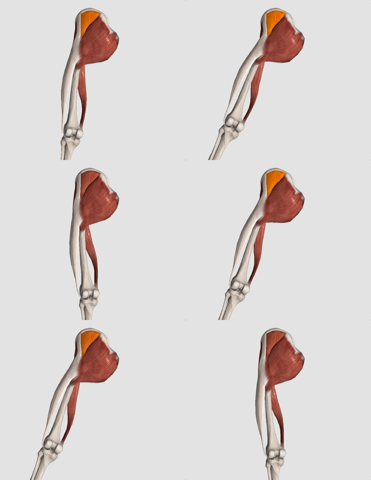
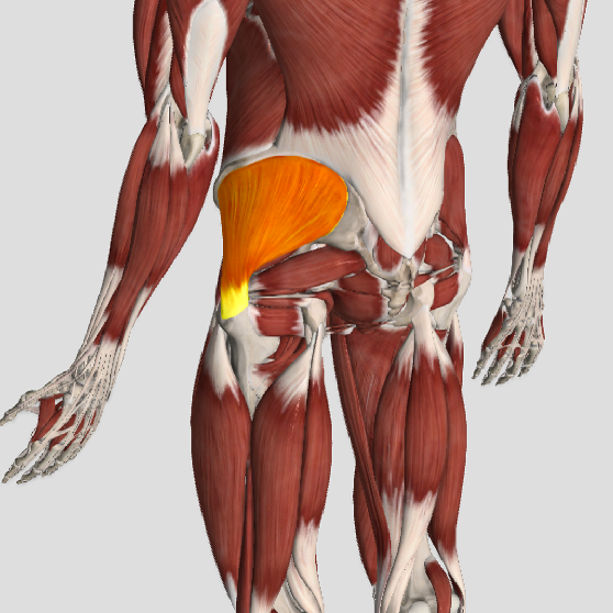
The gluteus medius is the principal hip abductor. When the hip is flexed, the muscle also assists the six deep hip external rotators (piriformis, gemelli, obturators, and quadratus femoris). The gluteus medius originates on the ilium just inferior to the iliac crest and inserts on the lateral and superior aspects of the greater trochanter. While the principal declared action of the gluteus medius is hip abduction, clinicians will appreciate its more valuable contribution as a dynamic stabilizer of the hip and pelvis- particularly during single leg stance activities like walking, running, and squatting. The gluteus medius contributes approximately 70% of the abduction force required to maintain pelvic leveling during single leg stance. The remainder comes predominantly from 2 muscles that insert onto the iliotibial band: the tensor fascia lata and upper gluteus maximus. Hip abductor strength is the single greatest contributor to lower extremity frontal plain alignment during activity. (6)
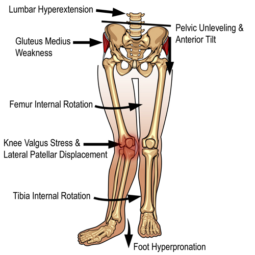
Incompetent hip abductors and/or external rotators allows for excessive adduction and internal rotation of the thigh during single leg stance activities. This leads to a cascade of biomechanical problems, including pelvic drop, excessive hip adduction, excessive femoral internal rotation, valgus knee stress, and internal tibial rotation. (1,7-12)
References
1. Szu-Ping Lee, Powers C. Description of a Weight-Bearing Method to Assess Hip Abductor and External Rotator Muscle Performance. JOSPT. Volume 43, Issue 6
2. Crossley KM, Zhang WJ, Schache AG, Bryant A, Cowan SM. Performance on the single-leg squat task indicates hip abductor muscle function. Am J Sports Med. 2011;39:866-873.
3. Presswood L, Cronin J, Keogh JWL, Whatman C. Gluteus medius: applied anatomy, dysfunction, assessment, and progressive strengthening. Strength Cond J. 2008;30:41-53.
4. Sled EA, Khoja L, Deluzio KJ, Olney SJ, Culham EG. Effect of a home program of hip abductor ex- ercises on knee joint loading, strength, function, and pain in people with knee osteoarthritis: a clinical trial. Phys Ther. 2010;90:895-904.
5. van der Krogt MM, Delp SL, Schwartz MH How robust is human gait to muscle weakness? Gait Posture. 2012 Feb 29.
6. Laurie Stickler, Margaret Finley, Heather Gulgin Relationship between hip and core strength and frontal plane alignment during a single leg squat Physical Therapy in Sport Available online 2 June 2014
7. Ireland ML, Willson JD, Ballantyne BT, Davis
IM. Hip strength in females with and without patellofemoral pain. J Orthop Sports Phys Ther. 2003;33:671-676.
8. Noehren B, Davis I, Hamill J. ASB clinical biome- chanics award winner 2006: prospective study of the biomechanical factors associated with iliotib- ial band syndrome. Clin Biomech (Bristol, Avon). 2007;22:951-956.
9. Powers CM. The influence of abnormal hip me- chanics on knee injury: a biomechanical perspec- tive. J Orthop Sports Phys Ther. 2010;40:42-51.
10. Powers CM. The influence of altered lower- extremity kinematics on patellofemoral joint dysfunction: a theoretical perspective. J Orthop Sports Phys Ther. 2003;33:639-646.
11. Sigward SM, Powers CM. Loading characteristics of females exhibiting excessive valgus moments during cutting. Clin Biomech (Bristol, Avon). 2007;22:827-833
12. Souza RB, Powers CM. Differences in hip kine- matics, muscle strength, and muscle activation between subjects with and without patellofemo- ral pain. J Orthop Sports Phys Ther. 2009;39:12- 19.
Patellofemoral pain syndrome (PFPS), also called “Runners Knee”, describes the symptom complex of knee discomfort, swelling, or crepitus that results from excessive or imbalanced forces acting on the joint. It is the most common cause of knee pain in the general population, affecting an estimated 25% of adults.
PFPS is most commonly related to lateral tracking of the patella. The patella has a natural tendency to migrate laterally due to the pull of the quadriceps and the slight natural valgus of the lower extremity. A new study in the Journal of Sports Medicine (1) provides additional confirmation that when managing patellofemoral pain syndrome, clinicians must address two critical yet often overlooked issues.
This study concludes that PFPS and dynamic knee valgus do not arise primarily from knee dysfunction, rather from hip abductor/ external rotator weakness and/or foot hyperpronation.
“The most effective intervention programs included exercises targeting the hip external rotator and abductor muscles and knee extensor muscles.” and “PFPS patients with foot abnormalities, such as those with increased rearfoot eversion or pes pronatus, may benefit the most from foot orthotics.”
Since gluteus medius and VMO weakness are key factors in the development of PFPS, strengthening exercises that target those muscles prove most effective. Stabilization exercises may include pillow push (push the back of your knee into a pillow for 5-6 seconds), supine heel slide, terminal knee (short-arc) extension, clam, glut bridge, semi-stiff deadlift, posterior lunge, and monster walk.
Myofascial release and stretching should be directed at hypertonic muscles, including the TFL, gastroc, soleus, hamstring, piriformis, hip rotators, and psoas. Myofascial release or IASTM may be appropriate for tightness in the iliotibial band, vastus lateralis, posterior hip capsule, and lateral knee retinaculum.
Manipulation may be necessary for restrictions in the lumbosacral and lower extremity joints. Hypermobility is common in the ipsilateral SI joint with restrictions present contralaterally. Evidence has shown that patellar tracking braces, i.e. BioSkin® or PatellaPro®, may lead to better outcomes.
Lifestyle modification may be necessary to reduce pain-provoking endeavors, especially running, jumping and other activities that induce a valgus stress. Athletes should avoid allowing their knee to cross in front of their toes while squatting. Arch supports or custom orthotics may be necessary to correct hyperpronation. Runners should avoid cross-over gaits and change shoes every 250 to 500 miles.
.
.
.
.
.
.
.
#pain
#kneepain
#chiropractic
#fitness
#sports
#wellness
#fitness
#healthyliving
#healthychoices
#Winnipeg
References
1. Petersen W, Rembitzki I, Liebau C. Patellofemoral pain in athletes. Open Access Journal of Sports Medicine. 2017;8:143-15



 Your low back relies on discs and other ligaments for support. “Discogenic Low Back Pain” develops when these tissues are placed under excessive stress, much like a rope that frays when it is stretched beyond its normal capacity. Most commonly, disc pain is not the result of any single event, but rather from repeated overloading. Your lumbar discs generally manage small isolated stressors quite well, but repetitive challenges lead to injury in much the same way that constantly bending a piece of copper wire will cause it to break. Examples of these stressors include: bad postures, sedentary lifestyles, poor fitting workstations, repetitive movements, improper lifting, or being overweight.