Fascial planes.

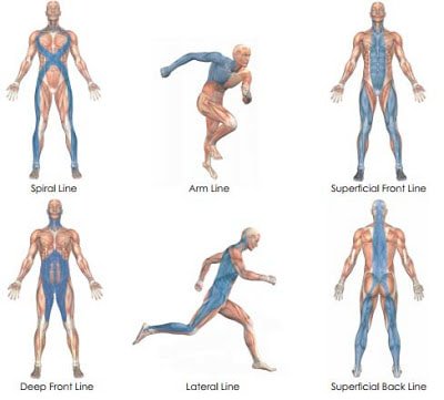
fascial connective tissue runs throughout all the bodies layers. It gives the body shape and function. Fascial litterally connects every part of your together. During movement when one part of the body moves, the body as a whole responds. Functionally the only tissue that can facilitate such responsiveness is the connective tissue. When one area of the body gets restricted or damaged, the body as a whole can become dysfunctional by setting up compensation patterns.

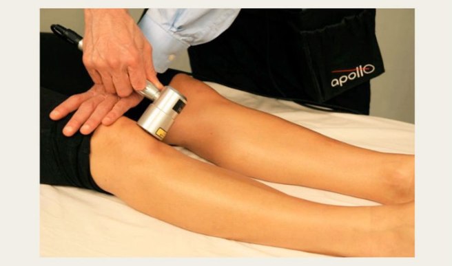
How Does Laser Work? 

Over 2000 published research studies demonstrate:

• Laser therapy improves blood flow and lymphatic drainage
• Laser therapy has a strengthening effect on tissue repair
• It is an effective means of relief for many pain syndromes
• It can improve immune response
• Enhanced nerve regeneration & function
• Increased microcirculation & vasodilation
• Increased lymphatic flow
• Increased collagen production
• Increases the speed, quality and tensile strength of tissue repair
• Reduced inflammation
• Enhanced angiogenesis (creation of new blood vessels)


Call us today for more information about Class IV laser therapy and how it can help you!


knee-apollo-image-716


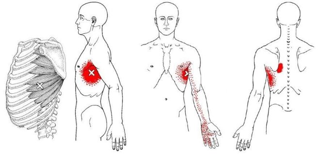
Trigger points in the serratus anterior.

The serratus anterior muscle is responsible for scapular and rib movement and stability. This muscle can become overloaded from poor posture of the shoulders, as well as heavy breathing from exertion. Trigger points in this muscle will cause pain to be felt under the armpit into the shoulder blade area, and down the arm into the fingers.

Trigger point referral patterns.

Myofascial trigger points form in a muscle due to overload stress. A portion of muscle fibers lock up into a knot. Once formed these points will irritate sensory nerves that are in proximity to the knot. When this happens,

trigger points have the capacity to refer pain along specific distributions or patterns that are well mapped out. sometimes pain may be felt at a great distance away from the actual point itself.

Trigger points.

Trigger points are knots of contracted muscle or connective tissue that form as a result of overload stress. Once formed these points will produce pain, refered pain, weakness, and stiffness. Trigger points can also mimic other conditions such as Carple tunnel syndrome and sciatica. Trigger points will on go away on their own, they must be manually released.

Trigger points in the trapezius muscle.

The trapezius muscle is a large diamond shaped muscle located in your back. This muscle is often overloaded due to poor sitting posture or excessive exercise. When this occurs trigger points will form. These points can cause back, neck, and shoulder pain. Trigger points in the upper traps are a leading cause of headache.

Trigger points in the gluteus Maximus muscle.

The gluteus Maximus muscle makes up your buttock. It’s is a powerful hip extensor and thus used heavily during activities such as walking, running, and climbing up stairs. Most atheletes abuse this muscle. When overworked trigger points will form, and these points will cause pain to be felt in the hip, sacrum and the as well as deep in the gluteal area. Litterally a pain in the butt!! Trigger points won’t release on their own and require interventions like trigger point massage.

MRI Truths & Myths

Arthritis

Low back pain is a very common complaint. In fact, it’s the #1 reason for doctor visits in the United States! The economic burden of LBP on the working class is astronomical. Most people can’t afford to be off work for one day, much less a week, month, or more! Because of the popularity of hospital-based TV dramas over the past two decades, many people think getting an MRI of their back can help their doctor fix their lower back problem. Is this a good idea? Let’s take a look!

Patients will often bring in a CD that has an MRI of their lower back to a doctor of chiropractic and ask the ultimate question, “….can you fix me?” Or, worse, “…I think I need surgery.” Sure, it’s quite amazing how an MRI can “slice” through the spine and show bone, soft tissues, disks, muscles, nerves, the spinal cord, and more! Since the low back bears approximately 2/3 of our body’s weight, you can frequently find MANY ABNORMALITIES in a person over 40-50 years old. In fact, it would be quite odd NOT to see things like disk degeneration, disk bulges, joint arthritis, spur formation, etc.!

Hence, the “downside” of having ALL this information is the struggle to determine which finding on the MRI has clinical significance. In other words, where is the LBP coming from? Is it that degenerative disk, bulged disk, herniated disk, or the narrowed canal where the nerve travels? Interestingly, in a recent review of more than 3,200 cases of acute low back pain, those who had an MRI scan performed earlier in their care had a WORSE outcome, more surgery, and higher costs compared with those who didn’t succumb to the temptation of requesting an MRI!

This is not to say MRI, CT scans, and x-rays are not important, as they effectively show conditions like subtle fractures and dangerous conditions like cancer. But for LBP, MRI is often misleading. This is because the primary cause of LBP is “functional” NOT “structural,” so it’s EASY to get railroaded into thinking whatever shows up on that MRI has to be the problem.

Here is how we know this, when we take 1,000 people WITHOUT low back pain between ages 30 and 60 (male or female) and perform an MRI on their lower back, we will find up to 53% will have PAINLESS disk bulges in one or more lumbar disks. Moreover, we will find up to 30% will have partial disk herniations, and up to 18% will have an extruded disk (one that has herniated ALL the way out). Yet, these people are PAIN FREE and never knew they had disk “derangement” (since they have no LBP). When combining all of these possible disk problems together, several studies report that between 57% and 64% of the general population has some type of disk problem without ANY BACK PAIN!

Hence, when a patient with a simple sprain/strain and localized LBP presents with an MRI showing a disk problem, it usually ONLY CONFUSES the patient (and frequently the doctor), as that disk problem is usually not the problem causing the pain!  So DON’T have an MRI UNLESS a surgical treatment decision depends on its findings. That is weakness, numbness, and non-resolving LBP in spite of 4-6 weeks of non-surgical care or unless there is weakness in bowel or bladder control. Remember, the majority of back pain sufferers DO NOT need surgery!

We realize you have a choice in whom you consider for your health care provision and we sincerely appreciate your trust in choosing our service for those needs.  If you, a friend, or family member requires care for back pain, we would be honored to render our services.

Trigger point massage

Session Description


A treatment with Bryan is very user friendly. And, no, you don’t have to remove any clothing. However, bringing a t-shirt and a pair of shorts or sweats is recommended.


The first time you come for a treatment you will be asked to fill out a Client History form. Bryan will go over the information you provide, asking for more detail and discussing the type of pain you are having and its location.


The treatment itself involves locating the Trigger Points in the muscle or soft tissue and applying a deep focused pressure to the Point. This will reproduce the pain and the referral pattern that is characteristic of that pain.


The treatment will be uncomfortable at first, but as the Trigger Points release, the pain will decrease. The pressure will always be adjusted to your tolerance level. If, at any time, you feel too uncomfortable you can ask Bryan to ease off a bit.


Depending on your specific problem, Bryan may also use some stretching and / or range-of-motion techniques, as needed.


After treatment, it is usually recommended that the client apply moist heat to the area treated.


What is the current “best evidence” in WRD right now?

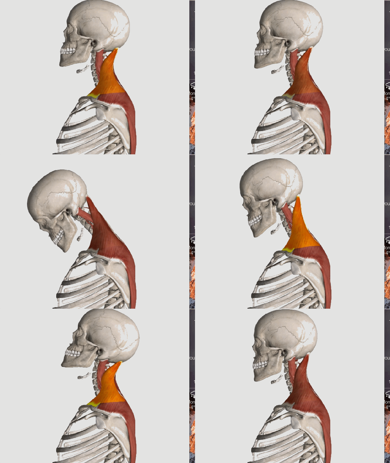
Up Trap Ext

Whiplash, or WAD (Whiplash Associated Disorders), refers to a neck injury where the normal range of motion is exceeded, resulting in injury to the soft-tissues (hopefully with no fractures) in the cervical region. There are a LOT of factors involved that enter into the degree of injury and length of healing time. Let’s take a closer look!

Picture the classic rear-end collision. The incident itself may be over within 300 milliseconds (msec), which is why it’s virtually impossible to brace yourself effectively for the crash as a typical voluntary muscle contraction takes two to three times longer (800-1000 msec) to accomplish.

In the first 50 msec, the force of the rear-end collision pushes the vehicle (and the torso of the body) forwards leaving the head behind so the cervical spine straightens out from its normal “C-shape” (or lordosis). By 75-100 msec, the lower part of the neck extends or becomes more C-shaped while the upper half flexes or moves in an opposite direction creating an “S” shape to the neck. Between 150-200msec, the whole neck hyper extends and the head may hit the head rest IF the headrest is positioned properly. In the last 200-300 msec, the head is propelled forwards into flexion in a “crack the whip” type of motion.

Injury to the neck may occur at various stages of this very fast process, and many factors determine the degree of injury such as a smaller car being hit by a larger car, the impact direction, the position of the head upon impact (worse if turned), if the neck is tall and slender vs. short and muscular, the angle and “springiness” of the seat back and relative position of the headrest, dry vs. wet/slippery pavement, and airbag deployment, just to name a few.

Some other factors that can predict recovery include: limited neck motion, the presence of neurological loss (nerve specific muscle weakness and/or numbness/tingling), high initial pain levels (>5/10 on a 0-10 scale), high disability scores on questionnaires, overly fearful of harming oneself with usual activity and/or work, depressive symptoms, post-traumatic stress, poor coping skills, headaches, back pain, widespread or whole body pain, dizziness, negative expectation of recovery, pending litigation, catastrophizing, age (older is worse), and poor pre-collision health (both mental and physical).

Research shows the best outcomes occur when patients are assured that most people fully recover and when patients stay active and working as much as possible. Studies have shown it’s best to avoid prolonged inactivity and cervical collars unless under a doctor’s orders. It’s also a good idea to gradually introduce exercises aimed at improving range of motion, postural endurance, and motor control provided doing so keeps the patient within reasonable pain boundaries. Chiropractic manipulation restores movement in fixed or stuck joints in the back and neck and has been found to help significantly with neck pain and headaches, particularly for patients involved in motor vehicle collisions. A doctor of chiropractic may also recommend using a cervical pillow, home traction, massage, and other therapies as part of the recovery process.

It is important to be aware that fear of normal activity and not engaging in usual activities and work can delay healing and promote chronic problems and long-term disability. It’s suggested patients avoid opioid medication use due to the addictive problems with such drugs. Ice and anti-inflammatory herbs or nutrients (like ginger, turmeric, and bioflavonoids) are safer options. Your doctor of chiropractic can guide you in this process!