Do you have CTS?

Carpal Tunnel

Carpal tunnel syndrome (CTS) can be an extremely painful and activity-limiting condition. It affects many people of all ages and genders, though women are affected more often than men. But how do you know if what you are suffering from is truly CTS or if it’s another condition that’s producing the symptoms in your hand or wrist?
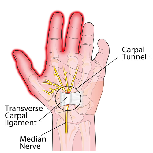
Carpal tunnel syndrome occurs when the median nerve is compressed as it passes through the wrist. However, the median nerve travels out of the neck, through the shoulder, elbow, and forearm before it passes through the wrist and into the hand. Pinching of the median nerve ANYWHERE along its course can give rise to the signs and symptoms of CTS including numbness, tingling, and/or pain into the hand and index, third, and thumb-side half of the fourth digits, and sometimes the thumb. If the pinch is significant enough, weakness can also occur. Sometimes the median nerve can become compressed at both the wrist and other body sites as it travels from the spinal cord to your hand, that’s why it’s important for a doctor to check for impingements along the entire course of the nerve.
But compression of the median nerve isn’t the only thing that can produce symptoms in the hand. Here are a couple of the more common conditions that are often confused with CTS:
1)  Ulnar neuropathy: This is pinching of the ulnar nerve (at the neck, shoulder, elbow, or wrist) but this gives rise to a similar numbness/tingling BUT into the pinky-side of the fourth and the fifth fingers (not the thumb-side of the hand). The most common pinch location is either at the neck or the inner elbow, the latter of which is called “cubital tunnel syndrome” or CuTS.
2)  Tendonitis: There are a total of nine tendons that pass through the carpal tunnel that help us grip or make a fist. Similarly, there are five main tendons on the back side of the hand that allow us to open our hands and spread our fingers. ANY of these tendons can get strained or torn, which results in swelling and pain as well as limited function BUT there is usually NO NUMBNESS/TINGLING!
3)  DeQuervain’s disease: This is really a tendonitis of an extensor tendon of the thumb and its synovial sheath that lubricates it resulting in a “tenosynovitis.” This creates pain with thumb movements, especially if you grasp your thumb in the palm of your hand and then bend your wrist sideways towards the pinky-side of the hand.
Chiropractors are well-trained to diagnose and treat patients with CTS. And if you don’t have CTS but another condition listed above, they can offer treatment (or a referral, if necessary) to help resolve it so you can return to your normal activities as soon as possible.

PFPS Cont. You want details?

Screen Shot 2017-10-31 at 11.15.32 AM.png

The muscles of the hip provide not only local stability, but also play an important role in spinal and lower extremity functional alignment. (1-4) While weakness in some hip muscles (hip extensors and knee extensors) is well tolerated, weakness or imbalance in others can have a profound effect on gait and biomechanical function throughout the lower half of the body. (5) Weakness of the hip abductors, particularly those that assist with external rotation, has the most significant impact on hip and lower extremity stability. (5,6)

Hip Abduction.gif

The gluteus medius is the principal hip abductor. When the hip is flexed, the muscle also assists the six deep hip external rotators (piriformis, gemelli, obturators, and quadratus femoris). The gluteus medius originates on the ilium just inferior to the iliac crest and inserts on the lateral and superior aspects of the greater trochanter. While the principal declared action of the gluteus medius is hip abduction, clinicians will appreciate its more valuable contribution as a dynamic stabilizer of the hip and pelvis- particularly during single leg stance activities like walking, running, and squatting. The gluteus medius contributes approximately 70% of the abduction force required to maintain pelvic leveling during single leg stance. The remainder comes predominantly from 2 muscles that insert onto the iliotibial band: the tensor fascia lata and upper gluteus maximus.  Hip abductor strength is the single greatest contributor to lower extremity frontal plain alignment during activity. (6)

Screen Shot 2017-10-31 at 11.12.00 AM

Incompetent hip abductors and/or external rotators allows for excessive adduction and internal rotation of the thigh during single leg stance activities. This leads to a cascade of biomechanical problems, including pelvic drop, excessive hip adduction, excessive femoral internal rotation, valgus knee stress, and internal tibial rotation. (1,7-12)


References
1. Szu-Ping Lee, Powers C. Description of a Weight-Bearing Method to Assess Hip Abductor and External Rotator Muscle Performance. JOSPT. Volume 43, Issue 6
2. Crossley KM, Zhang WJ, Schache AG, Bryant A, Cowan SM. Performance on the single-leg squat task indicates hip abductor muscle function. Am J Sports Med. 2011;39:866-873.
3. Presswood L, Cronin J, Keogh JWL, Whatman C. Gluteus medius: applied anatomy, dysfunction, assessment, and progressive strengthening. Strength Cond J. 2008;30:41-53.
4. Sled EA, Khoja L, Deluzio KJ, Olney SJ, Culham EG. Effect of a home program of hip abductor ex- ercises on knee joint loading, strength, function, and pain in people with knee osteoarthritis: a clinical trial. Phys Ther. 2010;90:895-904.
5. van der Krogt MM, Delp SL, Schwartz MH How robust is human gait to muscle weakness? Gait Posture. 2012 Feb 29.
6. Laurie Stickler, Margaret Finley, Heather Gulgin Relationship between hip and core strength and frontal plane alignment during a single leg squat Physical Therapy in Sport Available online 2 June 2014
7. Ireland ML, Willson JD, Ballantyne BT, Davis
IM. Hip strength in females with and without patellofemoral pain. J Orthop Sports Phys Ther. 2003;33:671-676.
8. Noehren B, Davis I, Hamill J. ASB clinical biome- chanics award winner 2006: prospective study of the biomechanical factors associated with iliotib- ial band syndrome. Clin Biomech (Bristol, Avon). 2007;22:951-956.
9. Powers CM. The influence of abnormal hip me- chanics on knee injury: a biomechanical perspec- tive. J Orthop Sports Phys Ther. 2010;40:42-51.
10. Powers CM. The influence of altered lower- extremity kinematics on patellofemoral joint dysfunction: a theoretical perspective. J Orthop Sports Phys Ther. 2003;33:639-646.
11. Sigward SM, Powers CM. Loading characteristics of females exhibiting excessive valgus moments during cutting. Clin Biomech (Bristol, Avon). 2007;22:827-833
12. Souza RB, Powers CM. Differences in hip kine- matics, muscle strength, and muscle activation between subjects with and without patellofemo- ral pain. J Orthop Sports Phys Ther. 2009;39:12- 19.

Does Fasting Work?

Exercise Tip


https://jamanetwork.com/journals/jamainternalmedicine/fullarticle/2517920#ioi160017r18

Some very interesting findings in this study. Over the course of two years participants who were part of the fasting group showed a weight loss of 10% on average as well as improved perceptions of quality of life, energy, sleep quality and libido.

Is fasting right for you? That depends and it is an idea that you and your health care professional should discuss as what works for some may not be right for you.

Patellofemoral Pain Syndrome

knock knee

Patellofemoral pain syndrome (PFPS), also called “Runners Knee”, describes the symptom complex of knee discomfort, swelling, or crepitus that results from excessive or imbalanced forces acting on the joint. It is the most common cause of knee pain in the general population, affecting an estimated 25% of adults.

PFPS is most commonly related to lateral tracking of the patella. The patella has a natural tendency to migrate laterally due to the pull of the quadriceps and the slight natural valgus of the lower extremity. A new study in the Journal of Sports Medicine (1) provides additional confirmation that when managing patellofemoral pain syndrome, clinicians must address two critical yet often overlooked issues.

This study concludes that PFPS and dynamic knee valgus do not arise primarily from knee dysfunction, rather from hip abductor/ external rotator weakness and/or foot hyperpronation.

“The most effective intervention programs included exercises targeting the hip external rotator and abductor muscles and knee extensor muscles.” and “PFPS patients with foot abnormalities, such as those with increased rearfoot eversion or pes pronatus, may benefit the most from foot orthotics.”

Since gluteus medius and VMO weakness are key factors in the development of PFPS, strengthening exercises that target those muscles prove most effective. Stabilization exercises may include pillow push (push the back of your knee into a pillow for 5-6 seconds), supine heel slide, terminal knee (short-arc) extension, clam, glut bridge, semi-stiff deadlift, posterior lunge, and monster walk.

Myofascial release and stretching should be directed at hypertonic muscles, including the TFL, gastroc, soleus, hamstring, piriformis, hip rotators, and psoas. Myofascial release or IASTM may be appropriate for tightness in the iliotibial band, vastus lateralis, posterior hip capsule, and lateral knee retinaculum.

Manipulation may be necessary for restrictions in the lumbosacral and lower extremity joints. Hypermobility is common in the ipsilateral SI joint with restrictions present contralaterally. Evidence has shown that patellar tracking braces, i.e. BioSkin® or PatellaPro®, may lead to better outcomes.

Lifestyle modification may be necessary to reduce pain-provoking endeavors, especially running, jumping and other activities that induce a valgus stress. Athletes should avoid allowing their knee to cross in front of their toes while squatting. Arch supports or custom orthotics may be necessary to correct hyperpronation. Runners should avoid cross-over gaits and change shoes every 250 to 500 miles.

.

.

.

.

.

.

.

#pain

#kneepain

#chiropractic

#fitness

#sports

#wellness

#fitness

#healthyliving

#healthychoices

#Winnipeg

References

1. Petersen W, Rembitzki I, Liebau C. Patellofemoral pain in athletes. Open Access Journal of Sports Medicine. 2017;8:143-15

Why Does My Back Hurt?

It’s been said that if you haven’t had back pain, just wait, because (statistically) some day you will! The following list is a list of “causes” that can be easily “fixed” to reduce your risk for a back pain episode.
1. MATTRESS: Which type of mattress is best? The “short answer”: there is no single mattress (style or type) for all people, primarily due to body type, size, gender, and what “feels good.” TRY laying on a variety of mattresses (for several minutes on your back and sides) and check out the difference between coiled, inner springs, foam (of different densities), air, waterbeds, etc. The thickness of a mattress can vary from 7 to 18 inches (~17-45 cm) deep. Avoid mattresses that feel like you’re sleeping in a hammock! A “good” mattress should maintain your natural spinal curves when lying on your sides or back (avoid stomach sleeping in most cases). Try placing a pillow between the knees and “hug” a pillow when side sleeping, as it can act like a “kick stand” and prevent you from rolling onto your stomach. If your budget is tight, you can “cheat” by placing a piece of plywood between the mattress and box spring as a short-term fix.
2. SHOES: Look at the bottom of your favorite pair of shoes and check out the “wear pattern.” If you have worn out soles or heels, you are way overdue for a new pair or a “re-sole” by your local shoe cobbler! If you work on your feet, then it’s even more important for both managing and preventing LBP!
3. DIET: A poor diet leads to obesity, which is a MAJOR cause of LBP. Consider the Paleo or Mediterranean Diet and STAY AWAY from fast food! Identify the two or three “food abuses” you have embraced and eliminate them – things with empty calories like soda, ice cream, chips… you get the picture! Keeping your BMI (Body Mass Index) between 20 and 25 is the goal! Positive “side-effects” include increased longevity, better overall health, and an improved quality of life!
4. EXERCISE: The most effective self-help approach to LBP management is exercise. Studies show those who exercise regularly hurt less, see doctors less, have a higher quality of life, and just feel better! This dovetails with diet in keeping your weight in check as well. Think of hamstring stretches and core strengthening as important LBP managers – USE PROPER TECHNIQUE AND FORM; YOUR DOCTOR OF CHIROPRACTIC CAN GUIDE YOU IN THIS PROCESS!
5. POSTURE: Another important “self-help” trick of the trade is to avoid sitting slumped over with an extreme forward head carriage positions. Remember that every inch your head pokes forwards places an additional ten pounds (~4.5 kg) of load on your upper back muscles to keep your head upright, and sitting slumped increases the load on your entire back!
We have only scratched the surface of some COMMON causes and/or contributors of back pain. Stay tuned next month as we continue this important conversation!
We realize you have a choice in whom you consider for your health care provision and we sincerely appreciate your trust in choosing our service for those needs.  If you, a friend, or family member requires care for back pain, we would be honored to render our services.