River East Chiropractic: Your Local Wellness Hub

Photo by Andrea Piacquadio on Pexels.com

Welcome to River East Chiropractic at 1187 Rothesay St!

We’re thrilled to introduce our new health and wellness clinic at 1187 Rothesay Street, right here in your neighbourhood. Whether you’re seeking pain relief, stress reduction, or a customized fitness plan, we’ve got you covered with a wide range of services designed to enhance your well-being.

Our Services

Here’s what you can expect when you visit us:

  • Chiropractic Care: Our skilled chiropractors provide effective treatments for back pain, headaches, sports injuries, and more. Let us help you get back to feeling your best.
  • Acupuncture: Whether you’re looking to relieve pain, reduce stress, or improve your overall health, our acupuncture services offer a natural and time-tested solution.
  • Laser Therapy: Using advanced technology, our low-level laser therapy speeds up healing and reduces pain, particularly for conditions like arthritis, soft tissue injuries, and chronic pain.
  • Gym & Personal Training: Our on-site gym offers top-notch equipment and personalized fitness plans from our experienced trainers to help you reach your health and fitness goals.
  • Free Child Care During Treatment: We understand how challenging it can be to focus on your health with little ones in tow, so we offer free child care while you receive treatment.
  • Massage Therapy: Soothe your aching muscles with our RMT

Convenient Hours for Your Busy Schedule

We know that life can be hectic, which is why we offer evening appointments until 8pm on Mondays and Thursdays to fit your schedule. Plus, we’re open from 9 AM to 1 PM on Saturdays to make it easier for you to prioritize your health.

Get in Touch

We’d love to welcome you to River East Chiropractic! For more information or to book an appointment, visit us online at rivereastchiropractic.ca or give us a call at (204) 334-3334. Your journey to better health starts here.

What is CTS?

480px-Carpal_Tunnel_Syndrome

Carpal tunnel syndrome (CTS) is a very common condition. According to a report by the Bureau of Labor Statistics (BLS), CTS ranks SECOND among the major disabling diseases and illnesses in ALL private industries. The BLS states that workers with CTS may eventually have to give up their livelihood. They cite one study in which almost half of all CTS patients changed their jobs within 30 months following their diagnosis. Due to the controversy surrounding the issue of CTS and worker’s compensation, workers do not always receive compensation benefits.
The KEY to long-term cost containment associated with CTS is EARLY DIAGNOSIS and PREVENTION! The challenge is getting the worker to identify early symptoms and NOT feel intimidated to report them, which could then lead to prompt care and possibly job modifications, resulting in the best chance of preventing a more complicated and far more costly problem.
Because of the many factors that contribute to and/or cause CTS, there is similarly no one way to prevent it from happening. Treating any/all underlying medical condition(s) is important. Using simple common sense can go a long way to help minimize some of the risk factors that predispose a person to work-related CTS and other cumulative trauma disorders (CTDs).
For example, watching and mimicking veteran workers can be a great guide as to how to maximize efficient work methods while minimizing unnecessary stresses and strains. Other preventative “tricks” include learning how to adjust the work area, handle tools, or perform tasks that minimize hand/wrist strain. Maintaining proper posture and exercise programs to strengthen the fingers, hands, wrists, forearms, shoulders, and neck may also help prevent CTS.
Many companies have taken action to help prevent repetitive stress injuries. In one study, 84% of the companies surveyed reported that they were modifying equipment, tasks, and processes as part of a prevention effort; nearly 85% analyzed their workstations and jobs; and 79% purchased more ergonomic equipment. Unfortunately, there is NO EVIDENCE that any of these methods can completely protect a worker against CTS. Often, the best approach is to relocate the worker to a less repetitive job, but this is not always an option.
Doctors of chiropractic can observe the worker through a video or during a factory tour/visit and often identify ergonomic problems that can result in a low-cost, easy modification. Simple modifications coupled with quality care, patient education, and cooperation from BOTH the worker and the employer can typically help yield the best outcome for the CTS patient.

What is Whiplash? 

WRD 2

Whiplash is an injury to the soft-tissues of the neck often referred to as a sprain or strain. Because there are a unique set of symptoms associated with whiplash, doctors and researchers commonly use the term “whiplash associated disorders” or WAD to describe the condition.

WAD commonly occurs as a result of a car crash, but it can also result from a slip and fall, sports injury, a personal injury (such as an assault), and other traumatic causes. The tissues commonly involved include muscle tendons (“strain”), ligaments and joint capsules (“sprains”), disk injuries (tears, herniation), as well as brain injury or concussion—even without hitting the head!

Symptoms vary widely but often include neck pain, stiffness, tender muscles and connective tissue (myofascial pain), headache, dizziness, sensations such as burning, prickly, tingling, numbness, muscle weakness, and referred pain to the shoulder blade, mid-back, arm, head, or face. If concussion occurs, additional symptoms include cognitive problems, concentration loss, poor memory, anxiety/depression, nervousness/irritability, sleep disturbance, fatigue, and more!

Whiplash associated disorders can be broken down into three categories: WAD I includes symptoms without any significant examination findings; WAD II includes loss of cervical range of motion and evidence of soft-tissue damage; and WAD III includes WAD II elements with neurological loss—altered motor and/or sensory functions. There is a WAD IV which includes fracture, but this is less common and often excluded.

Treatment for WAD includes everything from doing nothing to intensive management from multiple disciplines—chiropractic, primary care, physical therapy, clinical psychology, pain management, and specialty services such as neurology, orthopedics, and more. The goal of treatment is to restore normal function and activity participation, as well as symptom management.

The prognosis of WAD is generally good as many will recover without residual problems within days to weeks, with most people recovering around three months after the injury. Unfortunately, some are not so lucky and have continued neck pain, stiffness, headache, and some develop post-concussive syndrome. The latter can affect cognition, memory, vision, and other brain functions. Generally speaking, the higher the WAD category, the worse the prognosis, although each case MUST be managed by its own unique characteristics. If the injury includes neurological loss (muscle strength and/or sensory dysfunction like numbness, tingling, burning, pressure), the prognosis is often worse.

Chiropractic care for the WAD patient can include manipulation, mobilization, and home-based exercises, as well as the use of anti-inflammatory herbs (ginger, turmeric, proteolysis enzymes (bromelain, papain), devil’s claw, boswellia extract, rutin, bioflavonoid, vitamin D, coenzyme Q10, etc.) and dietary modifications aimed at reducing inflammation and promoting healing.

* 83% of those patients involved in an MVA will suffer whiplash injury and 50% will be symptomatic at 1 year.
* 90% of patients with neurologic signs at onset may be symptomatic at 1 year.
* 25- 80% of patients who suffer a whiplash injury will experience late-onset dizziness
* Clinicians should be observant for radiographic signs of instability, including interspinous widening, vertebral subluxation, vertebral compression fracture, and loss of cervical lordosis.
* Horizontal displacement of greater than 3.5 mm or angular displacement of more than 11 degrees on flexion/extension views suggests instability.

Why Does My Back Hurt?

It’s been said that if you haven’t had back pain, just wait, because (statistically) some day you will! The following list is a list of “causes” that can be easily “fixed” to reduce your risk for a back pain episode.
1. MATTRESS: Which type of mattress is best? The “short answer”: there is no single mattress (style or type) for all people, primarily due to body type, size, gender, and what “feels good.” TRY laying on a variety of mattresses (for several minutes on your back and sides) and check out the difference between coiled, inner springs, foam (of different densities), air, waterbeds, etc. The thickness of a mattress can vary from 7 to 18 inches (~17-45 cm) deep. Avoid mattresses that feel like you’re sleeping in a hammock! A “good” mattress should maintain your natural spinal curves when lying on your sides or back (avoid stomach sleeping in most cases). Try placing a pillow between the knees and “hug” a pillow when side sleeping, as it can act like a “kick stand” and prevent you from rolling onto your stomach. If your budget is tight, you can “cheat” by placing a piece of plywood between the mattress and box spring as a short-term fix.
2. SHOES: Look at the bottom of your favorite pair of shoes and check out the “wear pattern.” If you have worn out soles or heels, you are way overdue for a new pair or a “re-sole” by your local shoe cobbler! If you work on your feet, then it’s even more important for both managing and preventing LBP!
3. DIET: A poor diet leads to obesity, which is a MAJOR cause of LBP. Consider the Paleo or Mediterranean Diet and STAY AWAY from fast food! Identify the two or three “food abuses” you have embraced and eliminate them – things with empty calories like soda, ice cream, chips… you get the picture! Keeping your BMI (Body Mass Index) between 20 and 25 is the goal! Positive “side-effects” include increased longevity, better overall health, and an improved quality of life!
4. EXERCISE: The most effective self-help approach to LBP management is exercise. Studies show those who exercise regularly hurt less, see doctors less, have a higher quality of life, and just feel better! This dovetails with diet in keeping your weight in check as well. Think of hamstring stretches and core strengthening as important LBP managers – USE PROPER TECHNIQUE AND FORM; YOUR DOCTOR OF CHIROPRACTIC CAN GUIDE YOU IN THIS PROCESS!
5. POSTURE: Another important “self-help” trick of the trade is to avoid sitting slumped over with an extreme forward head carriage positions. Remember that every inch your head pokes forwards places an additional ten pounds (~4.5 kg) of load on your upper back muscles to keep your head upright, and sitting slumped increases the load on your entire back!
We have only scratched the surface of some COMMON causes and/or contributors of back pain. Stay tuned next month as we continue this important conversation!
We realize you have a choice in whom you consider for your health care provision and we sincerely appreciate your trust in choosing our service for those needs.  If you, a friend, or family member requires care for back pain, we would be honored to render our services.

What the heck is a trigger point?

trigger-point-referred-pain-shutterstock_228843211

What is a Trigger Point?

Trigger Points (TP’s) are defined as a “hyper-irritable spot within a taut band of skeletal muscle. The spot is painful on compression and can evoke characteristic referred pain and autonomic phenomena.”1

Put into plain language, a TP is a painful knot in muscle tissue that can refer pain to other areas of the body. You have probably felt the characteristic achy pain and stiffness that TP’s produce, at some time in your life.

TP’s were first brought to the attention of the medical world by Dr. Janet G. Travell. Dr. Travell, physician to President John F. Kennedy, is the acknowledged Mother of Myofascial Trigger Points. In fact, “Trigger Point massage, the most effective modality used by massage therapists for the relief of pain, is based almost entirely on Dr. Travell’s insights.”2 Dr. Travell’s partner in her research was Dr. David G. Simons, a research scientist and aerospace physician.

Trigger Points are very common. In fact, Travell and Simons state that TP’s are responsible for, or associated with, 75% of pain complaints or conditions.1 With this kind of prevalence, it’s no wonder that TP’s are often referred to as the “scourge of mankind”.

Trigger Points can produce a wide variety of pain complaints. Some of the most common are migraine headaches, back pain, and pain and tingling into the extremities. They are usually responsible for most cases of achy deep pain that is hard to localize.

A TP will refer pain in a predictable pattern, based on its location in a given muscle. Also, since these spots are bundles of contracted muscle fibres, they can cause stiffness and a decreased range of motion. Chronic conditions with many TP’s can also cause general fatigue and malaise, as well as muscle weakness.

Trigger Points are remarkably easy to get, but the most common causes are

TP’s (black X) can refer pain to other areas (red)

Sudden overload of a muscle

  • Poor posture
  • Chronic frozen posture (e.g., from a desk job), and
  • Repetitive strain

Once in place, a TP can remain there for the remainder of your life unless an intervention takes place.

Trigger Points Not Well Known

With thousands of people dealing with chronic pain, and with TP’s being responsible for — or associated with — a high percentage of chronic pain, it is very disappointing to find that a large portion of doctors and other health care practitioners don’t know about TP’s and their symptoms.

Scientific research on TP’s dates back to the 1700’s. There are numerous medical texts and papers written on the subject.

But, it still has been largely overlooked by the health care field. This has led to needless frustration and suffering, as well as thousands of lost work hours and a poorer quality of life.

How Are Trigger Points Treated?

As nasty and troublesome as TP’s are, the treatment for them is surely straight-forward. A skilled practitioner will assess the individual’s pain complaint to determine the most likely location of the TP’s and then apply one of several therapeutic modalities, the most effective of which is a massage technique called “ischemic compression”.

Basically, the therapist will apply a firm, steady pressure to the TP, strong enough to reproduce the symptoms. The pressure will remain until the tissue softens and then the pressure will increase appropriately until the next barrier is felt. This pressure is continued until the referral pain has subsided and the TP is released. (Note: a full release of TP’s could take several sessions.)

Other effective modalities include dry needling (needle placed into the belly of the TP) or wet needling (injection into the TP). The use of moist heat and stretching prove effective, as well. The best practitioners for TP release are Massage Therapists, Physiotherapists, and Athletic Therapists. An educated individual can also apply ischemic compression to themselves, but should start out seeing one of the above therapists to become familiar with the modality and how to apply pressure safely.

1 Simons, D.G., Travell, D.G., & Simons, L.S. Travell and Simons’Myofascial Pain and Dysfunction: the Trigger Point Manual.

Vol. 1. 2nd ed. Lippincott, Williams, and Wilkins, 1999.

PFPS Cont. You want details?

Screen Shot 2017-10-31 at 11.15.32 AM.png

The muscles of the hip provide not only local stability, but also play an important role in spinal and lower extremity functional alignment. (1-4) While weakness in some hip muscles (hip extensors and knee extensors) is well tolerated, weakness or imbalance in others can have a profound effect on gait and biomechanical function throughout the lower half of the body. (5) Weakness of the hip abductors, particularly those that assist with external rotation, has the most significant impact on hip and lower extremity stability. (5,6)

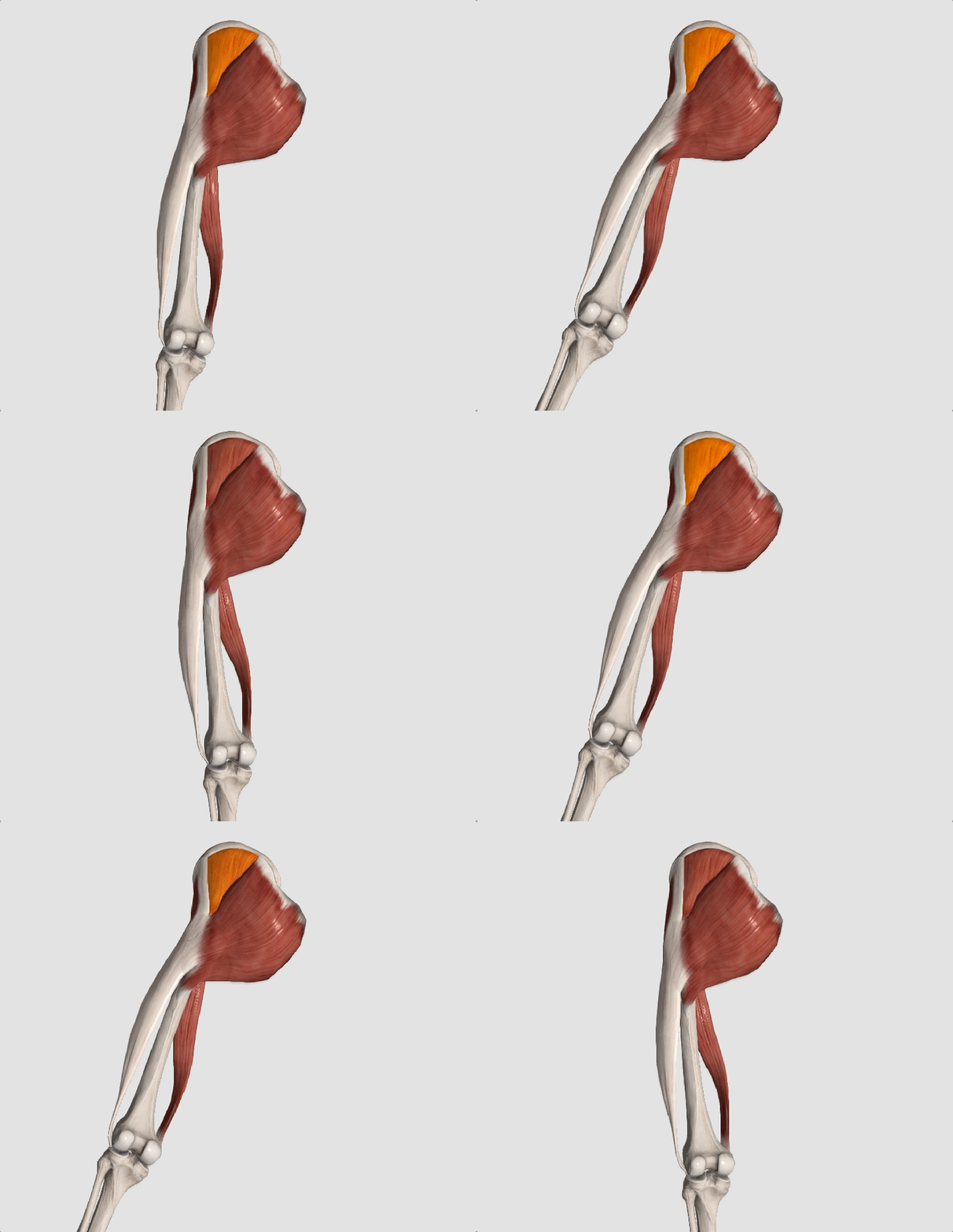
Hip Abduction.gif

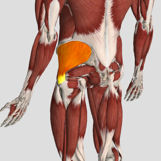
The gluteus medius is the principal hip abductor. When the hip is flexed, the muscle also assists the six deep hip external rotators (piriformis, gemelli, obturators, and quadratus femoris). The gluteus medius originates on the ilium just inferior to the iliac crest and inserts on the lateral and superior aspects of the greater trochanter. While the principal declared action of the gluteus medius is hip abduction, clinicians will appreciate its more valuable contribution as a dynamic stabilizer of the hip and pelvis- particularly during single leg stance activities like walking, running, and squatting. The gluteus medius contributes approximately 70% of the abduction force required to maintain pelvic leveling during single leg stance. The remainder comes predominantly from 2 muscles that insert onto the iliotibial band: the tensor fascia lata and upper gluteus maximus.  Hip abductor strength is the single greatest contributor to lower extremity frontal plain alignment during activity. (6)

Screen Shot 2017-10-31 at 11.12.00 AM

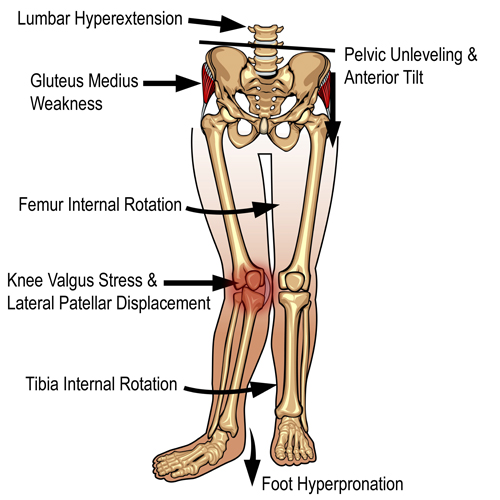
Incompetent hip abductors and/or external rotators allows for excessive adduction and internal rotation of the thigh during single leg stance activities. This leads to a cascade of biomechanical problems, including pelvic drop, excessive hip adduction, excessive femoral internal rotation, valgus knee stress, and internal tibial rotation. (1,7-12)


References
1. Szu-Ping Lee, Powers C. Description of a Weight-Bearing Method to Assess Hip Abductor and External Rotator Muscle Performance. JOSPT. Volume 43, Issue 6
2. Crossley KM, Zhang WJ, Schache AG, Bryant A, Cowan SM. Performance on the single-leg squat task indicates hip abductor muscle function. Am J Sports Med. 2011;39:866-873.
3. Presswood L, Cronin J, Keogh JWL, Whatman C. Gluteus medius: applied anatomy, dysfunction, assessment, and progressive strengthening. Strength Cond J. 2008;30:41-53.
4. Sled EA, Khoja L, Deluzio KJ, Olney SJ, Culham EG. Effect of a home program of hip abductor ex- ercises on knee joint loading, strength, function, and pain in people with knee osteoarthritis: a clinical trial. Phys Ther. 2010;90:895-904.
5. van der Krogt MM, Delp SL, Schwartz MH How robust is human gait to muscle weakness? Gait Posture. 2012 Feb 29.
6. Laurie Stickler, Margaret Finley, Heather Gulgin Relationship between hip and core strength and frontal plane alignment during a single leg squat Physical Therapy in Sport Available online 2 June 2014
7. Ireland ML, Willson JD, Ballantyne BT, Davis
IM. Hip strength in females with and without patellofemoral pain. J Orthop Sports Phys Ther. 2003;33:671-676.
8. Noehren B, Davis I, Hamill J. ASB clinical biome- chanics award winner 2006: prospective study of the biomechanical factors associated with iliotib- ial band syndrome. Clin Biomech (Bristol, Avon). 2007;22:951-956.
9. Powers CM. The influence of abnormal hip me- chanics on knee injury: a biomechanical perspec- tive. J Orthop Sports Phys Ther. 2010;40:42-51.
10. Powers CM. The influence of altered lower- extremity kinematics on patellofemoral joint dysfunction: a theoretical perspective. J Orthop Sports Phys Ther. 2003;33:639-646.
11. Sigward SM, Powers CM. Loading characteristics of females exhibiting excessive valgus moments during cutting. Clin Biomech (Bristol, Avon). 2007;22:827-833
12. Souza RB, Powers CM. Differences in hip kine- matics, muscle strength, and muscle activation between subjects with and without patellofemo- ral pain. J Orthop Sports Phys Ther. 2009;39:12- 19.

Patellofemoral Pain Syndrome

knock knee

Patellofemoral pain syndrome (PFPS), also called “Runners Knee”, describes the symptom complex of knee discomfort, swelling, or crepitus that results from excessive or imbalanced forces acting on the joint. It is the most common cause of knee pain in the general population, affecting an estimated 25% of adults.

PFPS is most commonly related to lateral tracking of the patella. The patella has a natural tendency to migrate laterally due to the pull of the quadriceps and the slight natural valgus of the lower extremity. A new study in the Journal of Sports Medicine (1) provides additional confirmation that when managing patellofemoral pain syndrome, clinicians must address two critical yet often overlooked issues.

This study concludes that PFPS and dynamic knee valgus do not arise primarily from knee dysfunction, rather from hip abductor/ external rotator weakness and/or foot hyperpronation.

“The most effective intervention programs included exercises targeting the hip external rotator and abductor muscles and knee extensor muscles.” and “PFPS patients with foot abnormalities, such as those with increased rearfoot eversion or pes pronatus, may benefit the most from foot orthotics.”

Since gluteus medius and VMO weakness are key factors in the development of PFPS, strengthening exercises that target those muscles prove most effective. Stabilization exercises may include pillow push (push the back of your knee into a pillow for 5-6 seconds), supine heel slide, terminal knee (short-arc) extension, clam, glut bridge, semi-stiff deadlift, posterior lunge, and monster walk.

Myofascial release and stretching should be directed at hypertonic muscles, including the TFL, gastroc, soleus, hamstring, piriformis, hip rotators, and psoas. Myofascial release or IASTM may be appropriate for tightness in the iliotibial band, vastus lateralis, posterior hip capsule, and lateral knee retinaculum.

Manipulation may be necessary for restrictions in the lumbosacral and lower extremity joints. Hypermobility is common in the ipsilateral SI joint with restrictions present contralaterally. Evidence has shown that patellar tracking braces, i.e. BioSkin® or PatellaPro®, may lead to better outcomes.

Lifestyle modification may be necessary to reduce pain-provoking endeavors, especially running, jumping and other activities that induce a valgus stress. Athletes should avoid allowing their knee to cross in front of their toes while squatting. Arch supports or custom orthotics may be necessary to correct hyperpronation. Runners should avoid cross-over gaits and change shoes every 250 to 500 miles.

.

.

.

.

.

.

.

#pain

#kneepain

#chiropractic

#fitness

#sports

#wellness

#fitness

#healthyliving

#healthychoices

#Winnipeg

References

1. Petersen W, Rembitzki I, Liebau C. Patellofemoral pain in athletes. Open Access Journal of Sports Medicine. 2017;8:143-15

Do you have CTS?

Carpal Tunnel

Carpal tunnel syndrome (CTS) can be an extremely painful and activity-limiting condition. It affects many people of all ages and genders, though women are affected more often than men. But how do you know if what you are suffering from is truly CTS or if it’s another condition that’s producing the symptoms in your hand or wrist?
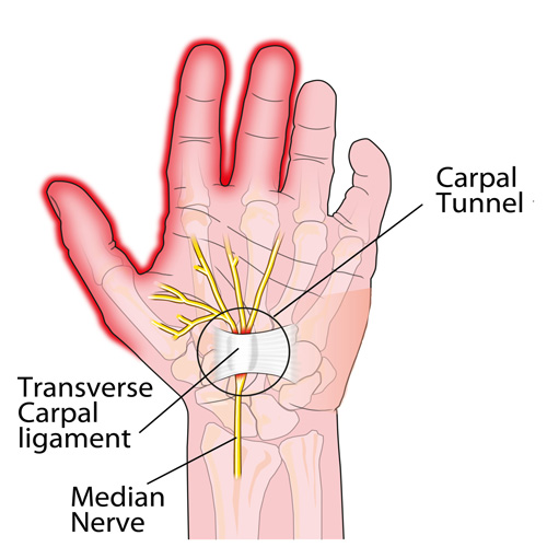
Carpal tunnel syndrome occurs when the median nerve is compressed as it passes through the wrist. However, the median nerve travels out of the neck, through the shoulder, elbow, and forearm before it passes through the wrist and into the hand. Pinching of the median nerve ANYWHERE along its course can give rise to the signs and symptoms of CTS including numbness, tingling, and/or pain into the hand and index, third, and thumb-side half of the fourth digits, and sometimes the thumb. If the pinch is significant enough, weakness can also occur. Sometimes the median nerve can become compressed at both the wrist and other body sites as it travels from the spinal cord to your hand, that’s why it’s important for a doctor to check for impingements along the entire course of the nerve.
 
But compression of the median nerve isn’t the only thing that can produce symptoms in the hand. Here are a couple of the more common conditions that are often confused with CTS:
1)  Ulnar neuropathy: This is pinching of the ulnar nerve (at the neck, shoulder, elbow, or wrist) but this gives rise to a similar numbness/tingling BUT into the pinky-side of the fourth and the fifth fingers (not the thumb-side of the hand). The most common pinch location is either at the neck or the inner elbow, the latter of which is called “cubital tunnel syndrome” or CuTS.
 
2)  Tendonitis: There are a total of nine tendons that pass through the carpal tunnel that help us grip or make a fist. Similarly, there are five main tendons on the back side of the hand that allow us to open our hands and spread our fingers. ANY of these tendons can get strained or torn, which results in swelling and pain as well as limited function BUT there is usually NO NUMBNESS/TINGLING!
 
3)  DeQuervain’s disease: This is really a tendonitis of an extensor tendon of the thumb and its synovial sheath that lubricates it resulting in a “tenosynovitis.” This creates pain with thumb movements, especially if you grasp your thumb in the palm of your hand and then bend your wrist sideways towards the pinky-side of the hand.
 
Chiropractors are well-trained to diagnose and treat patients with CTS. And if you don’t have CTS but another condition listed above, they can offer treatment (or a referral, if necessary) to help resolve it so you can return to your normal activities as soon as possible.