What is Whiplash?

Up Trap Ext

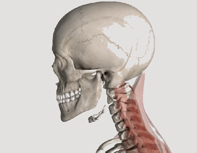
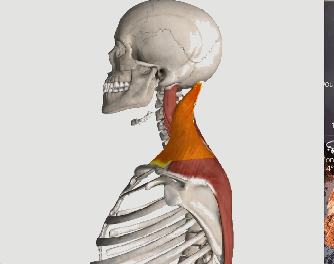
Whiplash is an injury to the soft-tissues of the neck often referred to as a sprain or strain. Because there are a unique set of symptoms associated with whiplash, doctors and researchers commonly use the term “whiplash associated disorders” or WAD to describe the condition.

WAD commonly occurs as a result of a car crash, but it can also result from a slip and fall, sports injury, a personal injury (such as an assault), and other traumatic causes. The tissues commonly involved include muscle tendons (“strain”), ligaments and joint capsules (“sprains”), disk injuries (tears, herniation), as well as brain injury or concussion—even without hitting the head!

Symptoms vary widely but often include neck pain, stiffness, tender muscles and connective tissue (myofascial pain), headache, dizziness, sensations such as burning, prickly, tingling, numbness, muscle weakness, and referred pain to the shoulder blade, mid-back, arm, head, or face. If concussion occurs, additional symptoms include cognitive problems, concentration loss, poor memory, anxiety/depression, nervousness/irritability, sleep disturbance, fatigue, and more!

Whiplash associated disorders can be broken down into three categories: WAD I includes symptoms without any significant examination findings; WAD II includes loss of cervical range of motion and evidence of soft-tissue damage; and WAD III includes WAD II elements with neurological loss—altered motor and/or sensory functions. There is a WAD IV which includes fracture, but this is less common and often excluded.

Treatment for WAD includes everything from doing nothing to intensive management from multiple disciplines—chiropractic, primary care, physical therapy, clinical psychology, pain management, and specialty services such as neurology, orthopedics, and more. The goal of treatment is to restore normal function and activity participation, as well as symptom management.

The prognosis of WAD is generally good as many will recover without residual problems within days to weeks, with most people recovering around three months after the injury. Unfortunately, some are not so lucky and have continued neck pain, stiffness, headache, and some develop post-concussive syndrome. The latter can affect cognition, memory, vision, and other brain functions. Generally speaking, the higher the WAD category, the worse the prognosis, although each case MUST be managed by its own unique characteristics. If the injury includes neurological loss (muscle strength and/or sensory dysfunction like numbness, tingling, burning, pressure), the prognosis is often worse.

Chiropractic care for the WAD patient can include manipulation, mobilization, and home-based exercises, as well as the use of anti-inflammatory herbs (ginger, turmeric, proteolysis enzymes (bromelain, papain), devil’s claw, boswellia extract, rutin, bioflavonoid, vitamin D, coenzyme Q10, etc.) and dietary modifications aimed at reducing inflammation and promoting healing.

* 83% of those patients involved in an MVA will suffer whiplash injury and 50% will be symptomatic at 1 year.
* 90% of patients with neurologic signs at onset may be symptomatic at 1 year.
* 25- 80% of patients who suffer a whiplash injury will experience late-onset dizziness
* Clinicians should be observant for radiographic signs of instability, including interspinous widening, vertebral subluxation, vertebral compression fracture, and loss of cervical lordosis.
* Horizontal displacement of greater than 3.5 mm or angular displacement of more than 11 degrees on flexion/extension views suggests instability

Whiplash and Your Posture

WRD 2.gif

Posture assessment is a key component of the chiropractic examination, and the posture of the head and neck is especially important for a patient recovering from a whiplash injury. Forward head carriage describes a state in which the head sits more forward on the shoulders than it should. In order for the muscles in the neck and shoulders to keep the head upright, they must work harder. This added strain can increase one’s risk for neck pain and headaches, which is why retraining posture is a key component to the management of neck pain and headaches in patients with or without a history of whiplash.

Forward head carriage also increases the distance between the back of the head and the headrest in the seated position, especially when the seat is reclined. In a rear-end collision, a gap greater than a half an inch between the head rest and the back of the head increases the probability of injury due to the greater distance the head can hyperextend as it rebounds backwards into the headrest.  This makes posture correction of forward head carriage an important aspect of treatment from both a preventative and curative perspective.

So this begs the question, can forward head carriage be corrected?  The simple answer is “yes!” One study evaluated the effects of a 16-week resistance and stretching program designed to address forward head posture and protracted shoulder positioning.

Researchers conducted the study in two separate secondary schools with 130 adolescents aged 15–17 years with forward head and protracted shoulder posture. The control group participated in a regular physical education (PE) program while the experimental group attended the same PE classes with the addition of specific exercises for posture correction. The research ream measured the teens’ shoulder head posture from the side using two different validated methods and tracked symptoms using a questionnaire. The results revealed a significant improvement in the shoulder and cervical angle in the experimental group that did not occur in the control group.

The conclusion of the study strongly supports that a 16-week resistance and stretching program is effective in decreasing forward head and protracted shoulder posture in adolescents.  This would suggest that a program such as this should be strongly considered in the regular curriculum of PE courses since this is such a common problem.

Doctors of chiropractic are trained to evaluate and manage forward head posture with shoulder protraction. This can prove beneficial in both the prevention as well as management of signs and symptoms associated with a whiplash injury.

What Causes Low Back Pain?

236

Low back pain (LBP) can arise from disks, nerves, joints, and the surrounding soft tissues.
To simplify the task of determining “What is causing my LBP?” the Quebec Task Force recommends that LBP be divided into three main categories: 1) Mechanical LBP; 2) Nerve root related back pain; and 3) Pathology or fracture. We will address the first two, as they are most commonly managed by chiropractors.
Making the proper diagnosis points your doctor in the right direction regarding treatment. It avoids time wasted by treating an unrelated condition, which runs the risk of increased chances of a poor and/or prolonged recovery. Low back pain is no exception! The “correct” diagnosis allows treatment to be focused and specific so that it will yield the best results.
Mechanical low back pain is the most commonly seen type of back pain, and it encompasses pain that arises from sprains, strains, facet and sacroiliac (SI) syndromes, and more. The main difference between this and nerve root-related LBP is the ABSENCE of a pinched nerve. Hence, pain typically does NOT radiate, and if it does, it rarely goes beyond the knee and normally does not cause weakness in the leg.
The mechanism of injury for both types of LBP can occur when a person does too much, maintains an awkward position for too long, or over bends, lifts, and/or twists. However, LBP can also occur “insidiously” or for seemingly no reason at all. However, in most cases, if one thinks hard enough, they can identify an event or a series of “micro-traumas” extending back in time that may be the “cause” of their current low back pain issues.
Nerve root-related LBP is less common but it is often more severe—as the pain associated with a pinched nerve is often very sharp, can radiate down a leg often to the foot, and cause numbness, tingling, and muscle weakness. The location of the weakness depends on which nerve is pinched. Think of the nerve as a wire to a light and the switch of the nerve is located in the back where it exits the spine. When the switch is turned on (the nerve is pinched), and the “light” turns on—possibly in the outer foot, middle foot, inner foot, or front, back or side of the thigh. In fact, there are seven nerves that innervate or “run” into our leg, so usually, a very specific location “lights up” in the limb.
Determining the cause of your low back pain helps your doctor of chiropractic determine which treatments may work best to alleviate your pain as well as where such treatments can be focused.

Do I have a tension headache? Or Migranes?

Cervicogenic

Most likely, everyone reading this article has had a headache at one time or another. The American Headache Society reports that nearly 40% of the population suffers from episodic headaches each year while 3% have chronic tension-type headaches. The United States Department of Health and Human Services estimates that 29.5 million Americans experience migraines, but tension headaches are more common than migraines at a frequency of 5 to 1. Knowing the difference between the two is important, as the proper diagnosis can guide treatment in the right direction.

TENSION HEADACHES: These typically result in a steady ache and tightness located in the neck, particularly at the base of the skull, which can irritate the upper cervical nerve roots resulting in radiating pain and/or numbness into the head. At times, the pain can reach the eyes but often stops at the top of the head. Common triggers include stress, muscle strain, or anxiety.

MIGRAINE HEADACHES: Migraines are often much more intense, severe, and sometimes incapacitating. They usually remain on one side of the head and are associated with nausea and/or vomiting. An “aura”, or a pre-headache warning, often comes with symptoms such as a bright flashing light, ringing or noise in the ears, a visual floater, and more. For migraine headaches, there is often a strong family history, which indicates genetics may play a role in their origin.

There are many causes for headaches. Commonly, they include lack of sleep and/or stress and they can also result from a recent injury—such as a car accident, and/or a sports injury—especially when accompanied by a concussion.

Certain things can “trigger” a migraine including caffeine, chocolate, citrus fruits, cured meats, dehydration, depression, diet (skipping meals), dried fish, dried fruit, exercise (excessive), eyestrain, fatigue (extreme), food additives (nitrites, nitrates, MSG), lights (bright, flickering, glare), menstruation, some medications, noise, nuts, odors, onions, altered sleep, stress, watching TV, red wine/alcohol, weather, etc.

Posture is also a very important consideration. A forward head carriage is not only related to headaches, but also neck and back pain. We’ve previously pointed out that every inch (2.54 cm) the average 12 pound head (5.44 kg) shifts forwards adds an EXTRA ten pounds (4.5 kg) of load on the neck and upper back muscles to keep the head upright.

So, what can be done for people who suffer from headaches? First, research shows chiropractic care is highly effective for patients with both types of headaches. Spinal manipulation, deep tissue release techniques, and nutritional counseling are common approaches utilized by chiropractors. Patients are also advised to use some of these self-management strategies at home as part of their treatment plan: the use of ice, self-trigger point therapy, exercise (especially strengthening the deep neck flexors), and nutritional supplements.

What Exercises Should I Do For Fibro?

outdoor-685417_960_720

Fibromyalgia (FM) is a very common, chronic condition where the patient describes “widespread pain” not limited to one area of the body. Hence, when addressing exercises for FM, one must consider the whole body. Perhaps one of the most important to consider is the squat.

If you think about it, we must squat every time we sit down, stand up, get in/out of our car, and in/out of bed. Even climbing and descending steps results in a squat-lunge type of movement.

The problem with squatting is that we frequently lose (or misuse) the proper way to do this when we’re in pain as the pain forces us to compensate, which can cause us to develop faulty movement patterns that can irritate our ankles, knees, hips, and spine (particularly the low back). In fact, performing a squatting exercise properly will strengthen the hips, which will help protect the spine, and also strengthens the glutel muscles, which can help you perform all the daily activities mentioned above.

The “BEST” type of squat is the free-standing squat. This is done by bending the ankles, knees, and hips while keeping a curve in the low back. The latter is accomplished by “…sticking the butt out” during the squat.

Do NOT allow the knees to drift beyond your toes! If you notice sounds coming from your knees they can be ignored IF they are not accompanied by pain. If you do have pain, try moving the foot of the painful knee about six inches (~15 cm) ahead of the other and don’t squat as far down. Move within “reasonable boundaries of pain” by staying away from positions that reproduce sharp, lancinating pain that lingers upon completion.

There are MANY exercises that help FM, but this one is particularly important!

The Best Diet For Fibromyalgia?

Anti-Inflam

Fibromyalgia (FM) and its cause remains a mystery, but most studies suggest that FM is NOT the result of a single event but rather a combination of many physical, chemical, and emotional stressors.

The question of the month regarding the BEST FM diet is intriguing since one might assume that the many causes should mean that there isn’t one dietary solution. But is that true? Could there be a “best diet” to help ease the symptoms from such a multi-faceted disorder?

Certainly, healthy eating is VERY important for ALL of us regardless of our current ailment(s). Obesity is rampant largely due to the fact that 60% of the calories consumed by the “typical” American center around eating highly inflaming food that include those rich in Sugar, Omega-6 oil, Flour, and Trans fats (“SOFT” foods, if you will!). Obesity has been cited as “an epidemic” largely due to kids and adults becoming too sedentary (watching TV, playing on electronic devices, etc.) and eating poorly.

Perhaps the BEST way to manage the pain associated with FM and to maintain a healthy BMI (Body Mass Index, or ratio between height and weight) is to substitute ANTI-INFLAMING foods for those that inflame (or SOFT foods).

You can simplify your diet by substituting OUT “fast foods” for fruits, vegetables, and lean meats. So there you have it. It’s that simple. The problem is making up your mind to change and then actually doing it. Once these two things take place, most everyone can easily “recalibrate” their caloric intake and easily adapt.

Not only have studies shown that chronic illnesses like heart disease, stroke, and diabetes significantly benefit by following this simple dietary shift, but so does pain arising from the musculoskeletal system! This is because the human body is made up largely of chemicals, and chemical shifts are constantly taking place when it moves. If you reach for an anti-inflammatory drug like ibuprofen or naproxen and it helps, it’s because you ARE inflamed and the drug reduces the pain associated with that inflammation. This is an indication that an anti-inflammatory diet WILL HELP as well (but without the negative side effects)!

The list of chronic conditions that result in muscle pain not only includes FM but also obesity, metabolic syndrome, and type II diabetes. Conditions like tension-type and migraine headaches, neck and back pain, disk herniation, and tendonopathies and MANY more ALL respond WELL to making this SIMPLE change in the diet. For more information on how to “DEFLAME,” visit http://www.deflame.com! It could be a potential “lifesaver!”

I get dizzy when I have a headache. Should I worry?

Vertigo_view_03,_Andels_Hotel_Łódź.jpg

Dizziness, neck pain, and headaches are very common symptoms that may or may not occur at the same time. Though this interrelationship exists, this month’s article will focus primarily on dizziness, particularly related to dizziness that occurs after standing.

First, it is important to point out that it is VERY common to be light headed or dizzy when standing up too fast, which is typically referred to as orthostatic hypotension (OH). OH is frequently referred to as a benign symptom, but new information may challenge this thought.

Let’s review what happens. When we are lying down, our heart does not have to work as hard as when we are upright; therefore, our blood pressure (BP) is usually lower while we lay in bed. When standing up, blood initially pools in the legs until an increase in blood pressure brings oxygen to the brain. This either resolves or prevents dizziness.

Orthostatic hypotension is defined as a blood pressure drop of >20 mm Hg systolic (the upper number—heart at FULL contraction), 10 mm Hg diastolic (lower number—heart at FULL rest), or both. This typically occurs within seconds to a few minutes after rising to a standing position.

There are two types of OH—delayed OH (DOH) where the onset of symptoms are not immediate but occur within three minutes of standing and “full” OH, which is more serious and occurs immediately upon rising. According to a 2016 study published in the prestigious journal Neurology, researchers reviewed the medical records of 165 people who had undergone autonomic nervous system testing for dizziness. The subjects averaged 59 years of age, and 48 were diagnosed with DOH, 42 with full OH, and 75 subjects didn’t have either condition.

During a ten-year follow-up, 54% of the DOH group progressed to OH, of which 31% developed a degenerative brain condition such as Parkinson’s disease or dementia. Those with initial DOH who also had diabetes were more likely to develop full OH vs. those without diabetes.

The early death rate in this 165 patient group was 29% for those with DOH, 64% with full OH, vs. 9% for those with neither diagnosed condition. The authors point out that those initially diagnosed with DOH who did NOT progress into full OH were given treatment that may have improved their blood pressure.

The authors state that a premature death might be avoided by having DOH and OH diagnosed and properly managed as early as possible. They point out that a prospective study is needed since this study only looked back at medical records of subjects who had nervous system testing performed at a specialized center, and therefore, these findings may not apply to the general population.

The value of this study is that this is the FIRST time a study described OH (or DOH) as a potentially serious condition with recommendations NOT to take OH/DOH lightly or view it as a benign condition. Since doctors see this a lot, a closer evaluation of the patient is in order.

Teens & Headaches? What?

Migrane

In 2016, researchers at Curtin University in Perth examined the seated posture and health data of 1,108 17-year olds in an effort to determine if any particular posture increased the risk of headaches/neck pain among late adolescents.

Among four posture subgroups—upright, intermediate, slumped thorax, and forward head—the researchers observed the following: participants who were slumped in their thoracic spine (mid-back region) and had their head forward when they sat were at higher odds of having mild, moderate, or severe depression; participants classified as having a more upright posture exercised more frequently, females were more likely to sit more upright than males; those who were overweight were more likely to sit with a forward neck posture; and taller people were more likely to sit upright.

While they found biopsychosocial factors like exercise frequency, depression, and body mass index (BMI) ARE associated with headaches and neck pain, their data did not suggest any one particular posture increased the risk of neck pain or headaches more than any other posture among the teenagers involved in the study.

This is noteworthy as studies with adults do indicate the risk for neck pain and headaches is greater in individuals with poor neck posture. In particular, postures such as forward head carriage, pinching a phone between the ear and shoulder, and prolonged neck/head rotation outside of neutral can all increase the risk of cervical disorders. This suggests that in younger bodies, the cause of neck pain and headaches may be multifactoral and not limited to just poor posture and that treatment must address all issues that may increase one’s risk for neck pain/headaches in order to reach a desired outcome.

The good news is that chiropractic has long embraced the biopsychosocial model of healthcare, looking at ALL factors that affect back and neck pain and quality of life. Through patient education, spinal manipulation, mobilization, exercise training, the use of modalities, and more, chiropractors can greatly help those struggling with neck pain and headaches!

“Other” Causes of Low Back Pain

316046269_1280x720

Between 80% and 90% of the general population will experience an episode of lower back pain (LBP) at least once during their lives. When it affects the young to middle-aged, we often use the term “non-specific LBP” to describe the condition. The geriatric population suffers from the “aging effects” of the spine—things like degenerative joint disease, degenerative disk disease, and spinal stenosis. Fractures caused by osteoporosis can also result in back pain.

The “good news” is that there are rare times when your doctor must consider a serious cause of LBP. That’s why he or she will ask about or check the following during your initial consultation: 1) Have you had bowel or bladder control problems? (This is to make sure a patient doesn’t have “cauda equina syndrome”—a very severely pinched nerve.) 2) Take a patient’s temperature and ask about any recent urinary or respiratory tract infections to rule out spinal infections. 3) To rule out cancer, a doctor may ask about a family or personal history of cancer, recent unexplained weight loss, LBP that won’t go away with time, or sleep interruptions that are out of the ordinary. 4) To rule out fractures, a doctor may also take x-rays if a patient is over age 70 regardless of trauma due to osteoporosis, over age 50 with minor trauma, and at any age with major trauma.
Once a doctor of chiropractic can rule out the “dangerous” causes of LBP, the “KEY” form of treatment is giving reassurance that LBP is manageable and advise LBP sufferers of ALL ages (especially the elderly) to KEEP MOVING! Of course, the speed at which we move depends on many things—first is safety, but perhaps more importantly is to NOT BECOME AFRAID to do things! As we age, we gradually fall out of shape and end up blaming our age for the inability to do simple normal activities. Regardless of age, we must GRADUALLY increase our activities to avoid the trap of sedentary habits resulting in deconditioning followed “fear avoidant behavior!”
Here are a few “surprising” reasons your back may be “killing you”: 1) You’re feeling down – That’s right, having “the blues” and more serious mood disorders, like depression, can make it more difficult to cope with pain. Also, depression often reduces the drive to exercise, may disturb sleep, and can affect dietary decisions—all of which are LBP contributors. 2) Your phone – Poor posture caused by holding a phone between your bent head and shoulder (get a headset!) or prolonged mobile phone use can increase your risk for spinal pain. 3) Your feet hurt, which makes you walk with an altered gait pattern, forcing compensatory movements up the “kinetic chain” leading to LBP. 4) Core muscle weakness, especially if you add to that a “pendulous abdomen” from being overweight—this is a recipe for disaster for LBP. 5) Tight short muscles such as hamstrings, hip rotator muscles, and/or tight hip joint capsules are common problems that contribute to LBP. Stretching exercises can REALLY help!

My Hip Hurts….

hip FAI

​Is it a labral tear? 

One of the structures that is frequently blamed for hip pain is called the labrum—the rubbery tissue that surrounds the socket helping to stabilize the hip joint. This tissue often wears and tears with age, but it can also be torn as a result of a trauma or sports-related injury.

The clinical significance of a labral tear of the hip is controversial, as these can be found in people who don’t have any pain at all. We know from studies of the intervertebral disks located in the lower back that disk herniation is often found in pain-free subjects—between 20-50% of the normal population.  In other words, the presence of abnormalities on an MRI is often poorly associated with patient symptoms, and the presence of a labral tear of the hip appears to be quite similar.

For instance, in a study of 45 volunteers (average age 38, range: 15–66 years old; 60% males) with no history of hip pain, symptoms, injury, or prior surgery, MRIs reviewed by three board-certified radiologists revealed a total of 73% of the hips had abnormalities, of which more than two-thirds were labral tears.

Another interesting study found an equal number of labral tears in a group of professional ballet dancers (both with and without hip pain) and in non-dancer control subjects of similar age and gender.

Another study showed that diagnostic blocks—a pain killer injected into the hip for diagnostic purposes to determine if it’s a pain generator—failed to offer relief for those with labral tears.

Doctors of chiropractic are trained to identify the origins of pain arising from the low back, pelvis, hip, and knee, all of which can mimic or produce hip symptoms.  Utilizing information derived from a careful history, examination, imaging (when appropriate), and functional tests, chiropractors can offer a nonsurgical, noninvasive, safe method of managing hip pain.