My Hip Hurts….

hip FAI

​Is it a labral tear? 

One of the structures that is frequently blamed for hip pain is called the labrum—the rubbery tissue that surrounds the socket helping to stabilize the hip joint. This tissue often wears and tears with age, but it can also be torn as a result of a trauma or sports-related injury.

The clinical significance of a labral tear of the hip is controversial, as these can be found in people who don’t have any pain at all. We know from studies of the intervertebral disks located in the lower back that disk herniation is often found in pain-free subjects—between 20-50% of the normal population.  In other words, the presence of abnormalities on an MRI is often poorly associated with patient symptoms, and the presence of a labral tear of the hip appears to be quite similar.

For instance, in a study of 45 volunteers (average age 38, range: 15–66 years old; 60% males) with no history of hip pain, symptoms, injury, or prior surgery, MRIs reviewed by three board-certified radiologists revealed a total of 73% of the hips had abnormalities, of which more than two-thirds were labral tears.

Another interesting study found an equal number of labral tears in a group of professional ballet dancers (both with and without hip pain) and in non-dancer control subjects of similar age and gender.

Another study showed that diagnostic blocks—a pain killer injected into the hip for diagnostic purposes to determine if it’s a pain generator—failed to offer relief for those with labral tears.

Doctors of chiropractic are trained to identify the origins of pain arising from the low back, pelvis, hip, and knee, all of which can mimic or produce hip symptoms.  Utilizing information derived from a careful history, examination, imaging (when appropriate), and functional tests, chiropractors can offer a nonsurgical, noninvasive, safe method of managing hip pain.

Carpel Tunnel in a nutshell

Here it is: carpal tunnel syndrome (CTS) in a nutshell!


WHAT: CTS is caused by an injury to the median nerve (MN) as it travels through the wrist.
WHERE: The eight small carpal bones and a ligament form a tunnel in which tendons and nerves pass through to reach the hand.
HOW: The MN gets pinched/irritated from repetitive stress.
WHY: The tunnel is tight as it includes the MN and nine rapidly moving muscle tendons!
PROGRESS: CTS usually starts slow and often progresses over weeks, months, even years.
SYMPTOMS: Pain, numbness, tingling, and/or weakness of the hand, sparing the little finger.
PROGNOSIS: CTS is easier to treat shortly after it starts, and waiting too long to seek care may lead to worse outcomes.
RISK FACTORS: 1) family history (genetics); 2) women are more likely to suffer from CTS than men; 3) age over 50; 4) manual jobs; 5) pregnancy; 6) conditions like diabetes, hypothyroid, rheumatoid arthritis (RA), osteoarthritis, autoimmune diseases (includes RA, certain types of thyroid disease), gout, kidney disease (especially dialysis patients), Down syndrome, amyloidosis, acromegaly, tumors on the median nerve; 7) medications (those that affect the immune system such as interleukin-2, possibly corticosteroids), anti-clotting drugs such as warfarin, hormone replacement, BCPs; 8) obesity; 9) smoking; 10) alcohol abuse; and 11) trauma/injuries (fractures, tendonitis).
TREATMENT: Ideally, treatment should begin as soon as possible after symptoms first start, but this RARELY occurs due its slow and gradual onset. Non-surgical care includes anti-inflammatory care (ice, anti-inflammatory nutrients—ginger, turmeric, bioflavonoids; NSAIDs like ibuprofen), wrist splinting (primarily at night), corticosteroid injections, job/ergonomic modifications, exercises (yoga, stretching, strengthening, and aerobic fitness), low level laser therapy, ice, acupuncture, and chiropractic care. Chiropractic care includes MANY of the above PLUS manual therapies applied to the neck, shoulder, arm, wrist, and hand.Carpal Tunnel

Condition Of The Month: Thoracic Joint Restriction

T Spine Joint

Your spine is made up of 24 bones stacked on top of each other with a soft “disc” between each segment to allow for flexibility. Normally, each joint in your spine should move freely and independently. When one or more of your spinal vertebra is slightly misaligned and restricted, we call this condition a “spinal segmental joint restriction”. A “thoracic joint restriction” means that this misalignment or restriction is located in your upper or mid-back region.

To help visualize this, imagine a normal spine functioning like a big spring moving freely in every direction. A spine with a joint restriction is like having a section of that spring welded together. The spring may still move as a whole, but a portion of it is no longer functioning.

Joint restrictions can develop in many ways. Sometimes they are brought on by an accident or an injury. Other times, they develop from repetitive strains or poor posture. Being overweight, smoking, strenuous work, and emotional stress can make you more susceptible to problems.

Restricted joints give rise to a self-perpetuating cycle of discomfort. Joint restriction causes swelling and inflammation, which triggers muscular guarding leading to more restriction. Since your spine functions as a unit, rather than as isolated pieces, a joint restriction in one area of your spine often causes “compensatory” problems in another. Think of this as a rowboat with multiple oarsmen on each side. When one rower quits, the others are placed under additional stress and can become overworked.

Joint restrictions most commonly cause local tenderness and discomfort. You may notice that your range of motion is limited. Movement may increase your discomfort. Pain from a restricted joint often trickles around your rib cage or up & down your spine. Be sure to tell your chiropractor if your symptoms include any chest pain, shortness of breath, unusual cough, indigestion or flu- like complaints.

Long-standing restrictions are thought to result in arthritis – much like the way a slightly misaligned wheel on your car causes premature wearing of your tire.

You should recognize that your problem is common and generally treatable. Chiropractic care has been shown to be the safest and most effective treatment for joint restrictions. Our office offers several tools to help ease your pain. To speed your recovery, you should avoid activities that increase your pain. Be sure to take frequent breaks from sedentary activity. Yoga has been shown to help back pain sufferers so consider joining a class or picking up a DVD.

What is CTS?

480px-Carpal_Tunnel_Syndrome

Carpal tunnel syndrome (CTS) is a very common condition. According to a report by the Bureau of Labor Statistics (BLS), CTS ranks SECOND among the major disabling diseases and illnesses in ALL private industries. The BLS states that workers with CTS may eventually have to give up their livelihood. They cite one study in which almost half of all CTS patients changed their jobs within 30 months following their diagnosis. Due to the controversy surrounding the issue of CTS and worker’s compensation, workers do not always receive compensation benefits.
The KEY to long-term cost containment associated with CTS is EARLY DIAGNOSIS and PREVENTION! The challenge is getting the worker to identify early symptoms and NOT feel intimidated to report them, which could then lead to prompt care and possibly job modifications, resulting in the best chance of preventing a more complicated and far more costly problem.
Because of the many factors that contribute to and/or cause CTS, there is similarly no one way to prevent it from happening. Treating any/all underlying medical condition(s) is important. Using simple common sense can go a long way to help minimize some of the risk factors that predispose a person to work-related CTS and other cumulative trauma disorders (CTDs).
For example, watching and mimicking veteran workers can be a great guide as to how to maximize efficient work methods while minimizing unnecessary stresses and strains. Other preventative “tricks” include learning how to adjust the work area, handle tools, or perform tasks that minimize hand/wrist strain. Maintaining proper posture and exercise programs to strengthen the fingers, hands, wrists, forearms, shoulders, and neck may also help prevent CTS.
Many companies have taken action to help prevent repetitive stress injuries. In one study, 84% of the companies surveyed reported that they were modifying equipment, tasks, and processes as part of a prevention effort; nearly 85% analyzed their workstations and jobs; and 79% purchased more ergonomic equipment. Unfortunately, there is NO EVIDENCE that any of these methods can completely protect a worker against CTS. Often, the best approach is to relocate the worker to a less repetitive job, but this is not always an option.
Doctors of chiropractic can observe the worker through a video or during a factory tour/visit and often identify ergonomic problems that can result in a low-cost, easy modification. Simple modifications coupled with quality care, patient education, and cooperation from BOTH the worker and the employer can typically help yield the best outcome for the CTS patient.

Why Does My Back Hurt?

It’s been said that if you haven’t had back pain, just wait, because (statistically) some day you will! The following list is a list of “causes” that can be easily “fixed” to reduce your risk for a back pain episode.
1. MATTRESS: Which type of mattress is best? The “short answer”: there is no single mattress (style or type) for all people, primarily due to body type, size, gender, and what “feels good.” TRY laying on a variety of mattresses (for several minutes on your back and sides) and check out the difference between coiled, inner springs, foam (of different densities), air, waterbeds, etc. The thickness of a mattress can vary from 7 to 18 inches (~17-45 cm) deep. Avoid mattresses that feel like you’re sleeping in a hammock! A “good” mattress should maintain your natural spinal curves when lying on your sides or back (avoid stomach sleeping in most cases). Try placing a pillow between the knees and “hug” a pillow when side sleeping, as it can act like a “kick stand” and prevent you from rolling onto your stomach. If your budget is tight, you can “cheat” by placing a piece of plywood between the mattress and box spring as a short-term fix.
2. SHOES: Look at the bottom of your favorite pair of shoes and check out the “wear pattern.” If you have worn out soles or heels, you are way overdue for a new pair or a “re-sole” by your local shoe cobbler! If you work on your feet, then it’s even more important for both managing and preventing LBP!
3. DIET: A poor diet leads to obesity, which is a MAJOR cause of LBP. Consider the Paleo or Mediterranean Diet and STAY AWAY from fast food! Identify the two or three “food abuses” you have embraced and eliminate them – things with empty calories like soda, ice cream, chips… you get the picture! Keeping your BMI (Body Mass Index) between 20 and 25 is the goal! Positive “side-effects” include increased longevity, better overall health, and an improved quality of life!
4. EXERCISE: The most effective self-help approach to LBP management is exercise. Studies show those who exercise regularly hurt less, see doctors less, have a higher quality of life, and just feel better! This dovetails with diet in keeping your weight in check as well. Think of hamstring stretches and core strengthening as important LBP managers – USE PROPER TECHNIQUE AND FORM; YOUR DOCTOR OF CHIROPRACTIC CAN GUIDE YOU IN THIS PROCESS!
5. POSTURE: Another important “self-help” trick of the trade is to avoid sitting slumped over with an extreme forward head carriage positions. Remember that every inch your head pokes forwards places an additional ten pounds (~4.5 kg) of load on your upper back muscles to keep your head upright, and sitting slumped increases the load on your entire back!
We have only scratched the surface of some COMMON causes and/or contributors of back pain. Stay tuned next month as we continue this important conversation!
We realize you have a choice in whom you consider for your health care provision and we sincerely appreciate your trust in choosing our service for those needs.  If you, a friend, or family member requires care for back pain, we would be honored to render our services.

What the heck is a trigger point?

trigger-point-referred-pain-shutterstock_228843211

What is a Trigger Point?

Trigger Points (TP’s) are defined as a “hyper-irritable spot within a taut band of skeletal muscle. The spot is painful on compression and can evoke characteristic referred pain and autonomic phenomena.”1

Put into plain language, a TP is a painful knot in muscle tissue that can refer pain to other areas of the body. You have probably felt the characteristic achy pain and stiffness that TP’s produce, at some time in your life.

TP’s were first brought to the attention of the medical world by Dr. Janet G. Travell. Dr. Travell, physician to President John F. Kennedy, is the acknowledged Mother of Myofascial Trigger Points. In fact, “Trigger Point massage, the most effective modality used by massage therapists for the relief of pain, is based almost entirely on Dr. Travell’s insights.”2 Dr. Travell’s partner in her research was Dr. David G. Simons, a research scientist and aerospace physician.

Trigger Points are very common. In fact, Travell and Simons state that TP’s are responsible for, or associated with, 75% of pain complaints or conditions.1 With this kind of prevalence, it’s no wonder that TP’s are often referred to as the “scourge of mankind”.

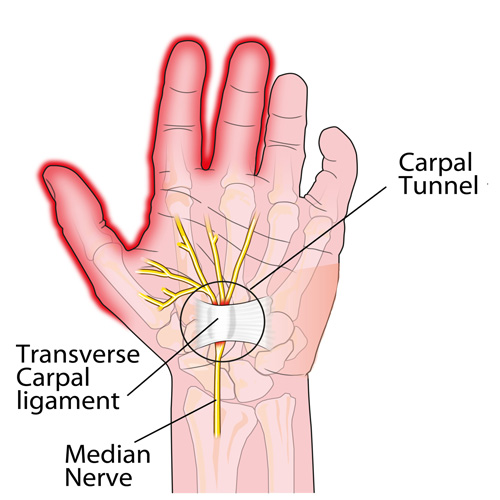
Trigger Points can produce a wide variety of pain complaints. Some of the most common are migraine headaches, back pain, and pain and tingling into the extremities. They are usually responsible for most cases of achy deep pain that is hard to localize.

A TP will refer pain in a predictable pattern, based on its location in a given muscle. Also, since these spots are bundles of contracted muscle fibres, they can cause stiffness and a decreased range of motion. Chronic conditions with many TP’s can also cause general fatigue and malaise, as well as muscle weakness.

Trigger Points are remarkably easy to get, but the most common causes are

TP’s (black X) can refer pain to other areas (red)

Sudden overload of a muscle

  • Poor posture
  • Chronic frozen posture (e.g., from a desk job), and
  • Repetitive strain

Once in place, a TP can remain there for the remainder of your life unless an intervention takes place.

Trigger Points Not Well Known

With thousands of people dealing with chronic pain, and with TP’s being responsible for — or associated with — a high percentage of chronic pain, it is very disappointing to find that a large portion of doctors and other health care practitioners don’t know about TP’s and their symptoms.

Scientific research on TP’s dates back to the 1700’s. There are numerous medical texts and papers written on the subject.

But, it still has been largely overlooked by the health care field. This has led to needless frustration and suffering, as well as thousands of lost work hours and a poorer quality of life.

How Are Trigger Points Treated?

As nasty and troublesome as TP’s are, the treatment for them is surely straight-forward. A skilled practitioner will assess the individual’s pain complaint to determine the most likely location of the TP’s and then apply one of several therapeutic modalities, the most effective of which is a massage technique called “ischemic compression”.

Basically, the therapist will apply a firm, steady pressure to the TP, strong enough to reproduce the symptoms. The pressure will remain until the tissue softens and then the pressure will increase appropriately until the next barrier is felt. This pressure is continued until the referral pain has subsided and the TP is released. (Note: a full release of TP’s could take several sessions.)

Other effective modalities include dry needling (needle placed into the belly of the TP) or wet needling (injection into the TP). The use of moist heat and stretching prove effective, as well. The best practitioners for TP release are Massage Therapists, Physiotherapists, and Athletic Therapists. An educated individual can also apply ischemic compression to themselves, but should start out seeing one of the above therapists to become familiar with the modality and how to apply pressure safely.

1 Simons, D.G., Travell, D.G., & Simons, L.S. Travell and Simons’Myofascial Pain and Dysfunction: the Trigger Point Manual.

Vol. 1. 2nd ed. Lippincott, Williams, and Wilkins, 1999.

PFPS Cont. You want details?

Screen Shot 2017-10-31 at 11.15.32 AM.png

The muscles of the hip provide not only local stability, but also play an important role in spinal and lower extremity functional alignment. (1-4) While weakness in some hip muscles (hip extensors and knee extensors) is well tolerated, weakness or imbalance in others can have a profound effect on gait and biomechanical function throughout the lower half of the body. (5) Weakness of the hip abductors, particularly those that assist with external rotation, has the most significant impact on hip and lower extremity stability. (5,6)

Hip Abduction.gif

The gluteus medius is the principal hip abductor. When the hip is flexed, the muscle also assists the six deep hip external rotators (piriformis, gemelli, obturators, and quadratus femoris). The gluteus medius originates on the ilium just inferior to the iliac crest and inserts on the lateral and superior aspects of the greater trochanter. While the principal declared action of the gluteus medius is hip abduction, clinicians will appreciate its more valuable contribution as a dynamic stabilizer of the hip and pelvis- particularly during single leg stance activities like walking, running, and squatting. The gluteus medius contributes approximately 70% of the abduction force required to maintain pelvic leveling during single leg stance. The remainder comes predominantly from 2 muscles that insert onto the iliotibial band: the tensor fascia lata and upper gluteus maximus.  Hip abductor strength is the single greatest contributor to lower extremity frontal plain alignment during activity. (6)

Screen Shot 2017-10-31 at 11.12.00 AM

Incompetent hip abductors and/or external rotators allows for excessive adduction and internal rotation of the thigh during single leg stance activities. This leads to a cascade of biomechanical problems, including pelvic drop, excessive hip adduction, excessive femoral internal rotation, valgus knee stress, and internal tibial rotation. (1,7-12)


References
1. Szu-Ping Lee, Powers C. Description of a Weight-Bearing Method to Assess Hip Abductor and External Rotator Muscle Performance. JOSPT. Volume 43, Issue 6
2. Crossley KM, Zhang WJ, Schache AG, Bryant A, Cowan SM. Performance on the single-leg squat task indicates hip abductor muscle function. Am J Sports Med. 2011;39:866-873.
3. Presswood L, Cronin J, Keogh JWL, Whatman C. Gluteus medius: applied anatomy, dysfunction, assessment, and progressive strengthening. Strength Cond J. 2008;30:41-53.
4. Sled EA, Khoja L, Deluzio KJ, Olney SJ, Culham EG. Effect of a home program of hip abductor ex- ercises on knee joint loading, strength, function, and pain in people with knee osteoarthritis: a clinical trial. Phys Ther. 2010;90:895-904.
5. van der Krogt MM, Delp SL, Schwartz MH How robust is human gait to muscle weakness? Gait Posture. 2012 Feb 29.
6. Laurie Stickler, Margaret Finley, Heather Gulgin Relationship between hip and core strength and frontal plane alignment during a single leg squat Physical Therapy in Sport Available online 2 June 2014
7. Ireland ML, Willson JD, Ballantyne BT, Davis
IM. Hip strength in females with and without patellofemoral pain. J Orthop Sports Phys Ther. 2003;33:671-676.
8. Noehren B, Davis I, Hamill J. ASB clinical biome- chanics award winner 2006: prospective study of the biomechanical factors associated with iliotib- ial band syndrome. Clin Biomech (Bristol, Avon). 2007;22:951-956.
9. Powers CM. The influence of abnormal hip me- chanics on knee injury: a biomechanical perspec- tive. J Orthop Sports Phys Ther. 2010;40:42-51.
10. Powers CM. The influence of altered lower- extremity kinematics on patellofemoral joint dysfunction: a theoretical perspective. J Orthop Sports Phys Ther. 2003;33:639-646.
11. Sigward SM, Powers CM. Loading characteristics of females exhibiting excessive valgus moments during cutting. Clin Biomech (Bristol, Avon). 2007;22:827-833
12. Souza RB, Powers CM. Differences in hip kine- matics, muscle strength, and muscle activation between subjects with and without patellofemo- ral pain. J Orthop Sports Phys Ther. 2009;39:12- 19.

Patellofemoral Pain Syndrome

knock knee

Patellofemoral pain syndrome (PFPS), also called “Runners Knee”, describes the symptom complex of knee discomfort, swelling, or crepitus that results from excessive or imbalanced forces acting on the joint. It is the most common cause of knee pain in the general population, affecting an estimated 25% of adults.

PFPS is most commonly related to lateral tracking of the patella. The patella has a natural tendency to migrate laterally due to the pull of the quadriceps and the slight natural valgus of the lower extremity. A new study in the Journal of Sports Medicine (1) provides additional confirmation that when managing patellofemoral pain syndrome, clinicians must address two critical yet often overlooked issues.

This study concludes that PFPS and dynamic knee valgus do not arise primarily from knee dysfunction, rather from hip abductor/ external rotator weakness and/or foot hyperpronation.

“The most effective intervention programs included exercises targeting the hip external rotator and abductor muscles and knee extensor muscles.” and “PFPS patients with foot abnormalities, such as those with increased rearfoot eversion or pes pronatus, may benefit the most from foot orthotics.”

Since gluteus medius and VMO weakness are key factors in the development of PFPS, strengthening exercises that target those muscles prove most effective. Stabilization exercises may include pillow push (push the back of your knee into a pillow for 5-6 seconds), supine heel slide, terminal knee (short-arc) extension, clam, glut bridge, semi-stiff deadlift, posterior lunge, and monster walk.

Myofascial release and stretching should be directed at hypertonic muscles, including the TFL, gastroc, soleus, hamstring, piriformis, hip rotators, and psoas. Myofascial release or IASTM may be appropriate for tightness in the iliotibial band, vastus lateralis, posterior hip capsule, and lateral knee retinaculum.

Manipulation may be necessary for restrictions in the lumbosacral and lower extremity joints. Hypermobility is common in the ipsilateral SI joint with restrictions present contralaterally. Evidence has shown that patellar tracking braces, i.e. BioSkin® or PatellaPro®, may lead to better outcomes.

Lifestyle modification may be necessary to reduce pain-provoking endeavors, especially running, jumping and other activities that induce a valgus stress. Athletes should avoid allowing their knee to cross in front of their toes while squatting. Arch supports or custom orthotics may be necessary to correct hyperpronation. Runners should avoid cross-over gaits and change shoes every 250 to 500 miles.

.

.

.

.

.

.

.

#pain

#kneepain

#chiropractic

#fitness

#sports

#wellness

#fitness

#healthyliving

#healthychoices

#Winnipeg

References

1. Petersen W, Rembitzki I, Liebau C. Patellofemoral pain in athletes. Open Access Journal of Sports Medicine. 2017;8:143-15

Chiro & Concussions

head-shot2

Whiplash Associated Disorders (WAD) is the appropriate terminology to use when addressing the myriad of symptoms that can occur as a result of a motor vehicle collision (MVC). In a recent publication in The Physician and Sports Medicine (Volume 43, Issue 3, 2015; 7/3/15 online:1-11), the article “The role of the cervical spine in post-concussive syndrome” takes a look at the neck when it’s injured in a car accident and how this relates to concussion.

It’s estimated about 3.8 million concussion injuries, also referred to as “mild traumatic brain injury” (mTBI), occur each year in the United States. Ironically, it’s one of the least understood injuries in the sports medicine and neuroscience communities. The GOOD NEWS is that concussion symptoms resolve within 7-10 days in the majority of cases; unfortunately, this isn’t the case with 10-15% of patients. Symptoms can last weeks, months, or even years in this group for which the term “post-concussive syndrome” (PCS) is used (defined as three or more symptoms lasting for four weeks as defined by the ICD-10) or three months following a minor head injury (as defined by the Diagnostic and Statistical Manual of Mental Disorders).

There have been significant advances in understanding what takes place in the acute phase of mTBI, but unfortunately, there is no clear physiological explanation for the chronic phase. Studies show the range of force to the head needed to cause concussion is between 60-160g (“g” = gravity) with 96.1g representing the highest predictive value in a football injury, whereas as little as 4.5g of neck acceleration can cause mild strain injury to the neck. In spite of this difference, the signs and symptoms reported by those injured in low-speed MVCs vs. football collisions are strikingly similar!

Research shows if an individual sustains an injury where the head is accelerated between 60-160g, it is HIGHLY likely that the tissues of the cervical spine (neck) have also reached their injury threshold of 4.5g. In a study that looked at hockey players, those who sustained a concussion also had WAD / neck injuries indicating that these injuries occur concurrently. Injuries to the neck in WAD include the same symptoms that occur in concussion including headache, dizziness/balance loss, nausea, visual and auditory problems, and cognitive dysfunction, just to name a few.

The paper concludes with five cases of PCS that responded well to a combination of active exercise/rehabilitation AND passive manual therapy (cervical spine manipulation). The favourable outcome supports the concept that the neck injury portion of WAD is a very important aspect to consider when treating patients with PCS!

This “link” between neck injury and concussion explains why chiropractic care is essential in the treatment of the concussion patient! This is especially true when the symptoms of concussion persist longer than one month!

We realize you have a choice in whom you consider for your health care provision and we sincerely appreciate your trust in choosing our service for those needs.  If you, a friend, or family member requires care for Whiplash, we would be honoured to render our services.