Neck Pain Reduction Strategies

Cervical Strain

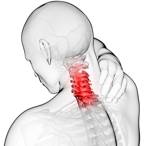
Neck pain is very common! According to one study, between 10-21% of the population will experience an episode of neck pain each year with a higher incidence rate among office workers. Between 33-65% will recover within one year, but most cases become “chronic, recurrent” meaning neck pain will come and go indefinitely. The more we can learn WHAT to do to prevent these episodes, the better.

1.  SLEEP: Use a cervical pillow so the NECK is fully supported during sleep. This keeps your head in alignment with your spine. Also, if possible, sleep on your back!

2. OFFICE: Position the computer screen so that it’s at or slightly below eye level and straight in front of you. The “KEY” point is that you feel comfortable with the height of the monitor. Keep your chin “tucked in” so the 10-11 pound (4.5-5 kg) weight of your head stays back over your shoulders—this will place less of a load on your upper back and neck muscles to hold your head upright! Set a timer on your cell phone to remind you to get up and move around every 30-60 minutes.

3. TELEPHONE: If you are using the phone a lot during the day, GET A HEADSET! If you are pinching the phone between your shoulder and ear, you WILL have neck problems!

4.  EXERCISE: Studies show people who are more physically active are less likely to report neck pain.

5.  NUTRITION: Search for information on the “anti-inflammatory diet.” It’s basically fruits, veggies, and lean meat, with a few other twists. Also, stay hydrated by drinking plenty of water each day.

6.  LIFT/CARRY: A heavy purse, brief case, or roller bag can really hurt your neck. Take ONLY what you need and put the rest in a secondary bag that stays in your car or where you can access it when needed. Switch to a backpack if possible vs. a heavy brief case.

7.  SELF-MASSAGE: Reach back and dig your fingers into your neck muscles and “work” the tight fibers back and forth until they loosen up. Roll your head over the top edge of a chair by sliding down until the top of the chair back rests in your neck. Search for the tight fibers and work them loose!

8.  WHIPLASH: If you are injured, DO NOT WAIT! Those who seek chiropractic care shortly after an accident have less long-term trouble!

PFPS Cont. You want details?

Screen Shot 2017-10-31 at 11.15.32 AM.png

The muscles of the hip provide not only local stability, but also play an important role in spinal and lower extremity functional alignment. (1-4) While weakness in some hip muscles (hip extensors and knee extensors) is well tolerated, weakness or imbalance in others can have a profound effect on gait and biomechanical function throughout the lower half of the body. (5) Weakness of the hip abductors, particularly those that assist with external rotation, has the most significant impact on hip and lower extremity stability. (5,6)

Hip Abduction.gif

The gluteus medius is the principal hip abductor. When the hip is flexed, the muscle also assists the six deep hip external rotators (piriformis, gemelli, obturators, and quadratus femoris). The gluteus medius originates on the ilium just inferior to the iliac crest and inserts on the lateral and superior aspects of the greater trochanter. While the principal declared action of the gluteus medius is hip abduction, clinicians will appreciate its more valuable contribution as a dynamic stabilizer of the hip and pelvis- particularly during single leg stance activities like walking, running, and squatting. The gluteus medius contributes approximately 70% of the abduction force required to maintain pelvic leveling during single leg stance. The remainder comes predominantly from 2 muscles that insert onto the iliotibial band: the tensor fascia lata and upper gluteus maximus.  Hip abductor strength is the single greatest contributor to lower extremity frontal plain alignment during activity. (6)

Screen Shot 2017-10-31 at 11.12.00 AM

Incompetent hip abductors and/or external rotators allows for excessive adduction and internal rotation of the thigh during single leg stance activities. This leads to a cascade of biomechanical problems, including pelvic drop, excessive hip adduction, excessive femoral internal rotation, valgus knee stress, and internal tibial rotation. (1,7-12)


References
1. Szu-Ping Lee, Powers C. Description of a Weight-Bearing Method to Assess Hip Abductor and External Rotator Muscle Performance. JOSPT. Volume 43, Issue 6
2. Crossley KM, Zhang WJ, Schache AG, Bryant A, Cowan SM. Performance on the single-leg squat task indicates hip abductor muscle function. Am J Sports Med. 2011;39:866-873.
3. Presswood L, Cronin J, Keogh JWL, Whatman C. Gluteus medius: applied anatomy, dysfunction, assessment, and progressive strengthening. Strength Cond J. 2008;30:41-53.
4. Sled EA, Khoja L, Deluzio KJ, Olney SJ, Culham EG. Effect of a home program of hip abductor ex- ercises on knee joint loading, strength, function, and pain in people with knee osteoarthritis: a clinical trial. Phys Ther. 2010;90:895-904.
5. van der Krogt MM, Delp SL, Schwartz MH How robust is human gait to muscle weakness? Gait Posture. 2012 Feb 29.
6. Laurie Stickler, Margaret Finley, Heather Gulgin Relationship between hip and core strength and frontal plane alignment during a single leg squat Physical Therapy in Sport Available online 2 June 2014
7. Ireland ML, Willson JD, Ballantyne BT, Davis
IM. Hip strength in females with and without patellofemoral pain. J Orthop Sports Phys Ther. 2003;33:671-676.
8. Noehren B, Davis I, Hamill J. ASB clinical biome- chanics award winner 2006: prospective study of the biomechanical factors associated with iliotib- ial band syndrome. Clin Biomech (Bristol, Avon). 2007;22:951-956.
9. Powers CM. The influence of abnormal hip me- chanics on knee injury: a biomechanical perspec- tive. J Orthop Sports Phys Ther. 2010;40:42-51.
10. Powers CM. The influence of altered lower- extremity kinematics on patellofemoral joint dysfunction: a theoretical perspective. J Orthop Sports Phys Ther. 2003;33:639-646.
11. Sigward SM, Powers CM. Loading characteristics of females exhibiting excessive valgus moments during cutting. Clin Biomech (Bristol, Avon). 2007;22:827-833
12. Souza RB, Powers CM. Differences in hip kine- matics, muscle strength, and muscle activation between subjects with and without patellofemo- ral pain. J Orthop Sports Phys Ther. 2009;39:12- 19.